

A multi-focus image fusion method of endoscope
A technology of image fusion and endoscopy, which is applied in the field of image processing, can solve the problems of poor imaging quality of endoscopy, achieve consistent image tone and brightness, eliminate plaque effect, and reduce the amount of calculation
Active Publication Date: 2022-06-28
THE AFFILIATED HOSPITAL OF QINGDAO UNIV
View PDF6 Cites 0 Cited by
- Summary
- Abstract
- Description
- Claims
- Application Information
AI Technical Summary
Problems solved by technology
[0007] Aiming at the technical problem of poor endoscope imaging quality caused by the defects of the image acquisition equipment itself or the poor image acquisition environment in the prior art, the present invention proposes an endoscope multi-focus image fusion method for improving endoscope acquisition output image quality
Method used
View moreImage
Smart ImageViewing Examples


Examples
Embodiment 1
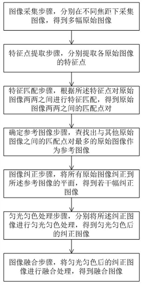
[0062] This embodiment proposes an endoscope multi-focus image fusion method, such as figure 1 shown, including:
[0063] In the image acquisition step, images are acquired at different focal lengths to obtain multiple original images.
[0064] First, the endoscope is used to collect images at different focusing distances, and the feature points of each image are extracted by the point feature extraction method to obtain the feature description of the feature points.
[0065] In the feature point extraction step, feature points of each original image are extracted respectively.
Login to View More PUM
 Abstract
The invention discloses an endoscope multi-focus image fusion method, which belongs to the technical field of image data processing, comprising: an image acquisition step, collecting images at different focal lengths to obtain multiple original images; a feature point extraction step, extracting each The feature points of the original image; the feature matching step is to obtain the matching point pairs between the original images; the step of determining the reference image is to find the reference image; the image correction step is to correct all the original images to the plane of the reference image to obtain several corrections image; the step of uniform light and color uniform processing, obtaining a corrected image after uniform light and color; the image fusion step, performing fusion processing on the corrected image after uniform light and color, to obtain a fusion image. In the endoscope multi-focus image fusion method of the present invention, an image including clear positions of all collected images is obtained by fusing multiple collected images with different focus distances, which is convenient for users to observe.
Description
technical field [0001] The invention belongs to the technical field of image processing, in particular to an endoscope multi-focus image fusion method. Background technique [0002] As a detection instrument based on image sensors, endoscopes can see lesions that cannot be displayed by X-rays, and are widely used in the medical field. Most of the existing endoscope photosensitive devices are CCD or CMOS, the imaging principle is pinhole imaging, and the clear range of imaging is limited by the depth of field. [0003] In addition, when imaging some organs of the human body, it is necessary to find specific pathological areas. However, because the human intestine is always in a state of peristalsis, the image resolution of the captured pathological areas will be extremely low, and even with Due to the peristalsis of the intestine, the pathological area cannot be kept in the middle of the image, which is not convenient for doctors to view. [0004] Due to the above-mentioned...
Claims
Login to View More Application Information
Patent Timeline
 Patent Type & Authority Patents(China)
IPC IPC(8): G06T5/50G06T5/00G06T7/90
CPCG06T5/50G06T5/007G06T7/90G06T2207/10068G06T2207/20221
Inventor 于腾波马金龙王辰陈进利付海涛王晓南王坤
Owner THE AFFILIATED HOSPITAL OF QINGDAO UNIV