Medical image-based bleeding area detection method and device, and storage medium

A medical imaging and area detection technology, applied in the field of medical imaging, can solve problems such as differences in conclusions, low efficiency and poor accuracy of cerebral hemorrhage, and achieve the effect of improving recognition accuracy and efficiency

Pending Publication Date: 2022-04-29
沈阳先进医疗设备技术孵化中心有限公司
View PDF0 Cites 0 Cited by
  • Summary
  • Abstract
  • Description
  • Claims
  • Application Information

AI Technical Summary

Problems solved by technology

The recognition results are affected by subjective factors. Doctors with different sets of data give different evaluations. The same doctor looks at the same set of data twice before and after, and the conclusions given will also be different.
At present, there are problems of low efficiency and poor accuracy in the identification of cerebral hemorrhage

Method used


Image

  • Medical image-based bleeding area detection method and device, and storage medium
  • Medical image-based bleeding area detection method and device, and storage medium
  • Medical image-based bleeding area detection method and device, and storage medium

Examples


Embodiment Construction

[0073] Hereinafter, the present application will be described in detail with reference to the drawings and embodiments. It should be noted that, in the case of no conflict, the embodiments in the present application and the features in the embodiments can be combined with each other.

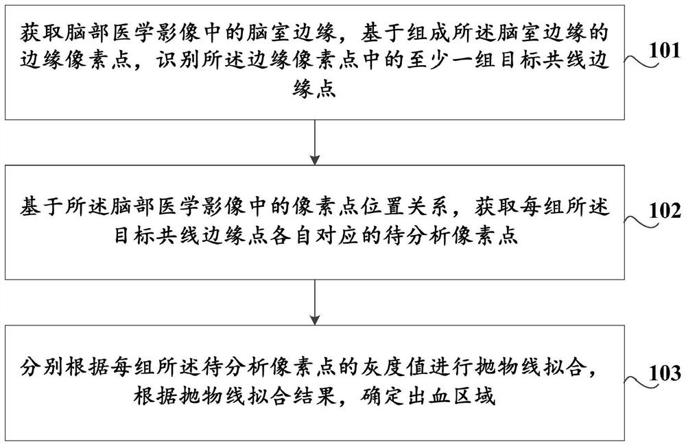
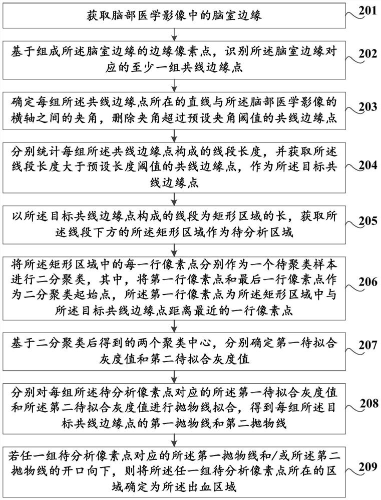
[0074] In this embodiment, a method for detecting bleeding areas based on medical images is provided, such as figure 1 As shown, the method includes:

[0075] Step 101, acquiring the ventricle edge in the brain medical image, and identifying at least one group of target collinear edge points in the edge pixel points based on the edge pixel points forming the ventricle edge;

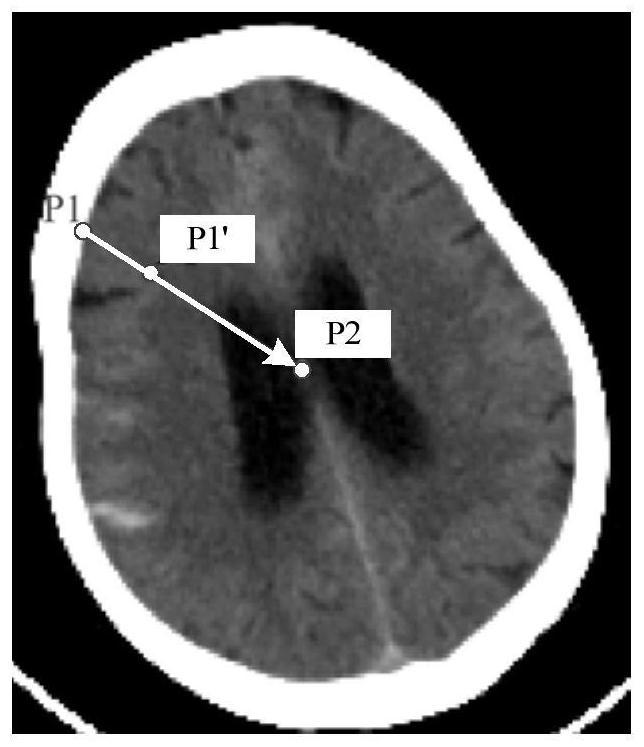
[0076] In the embodiment of the present application, it is possible to identify whether the patient has cerebral hemorrhage by analyzing the medical image of the patient's brain. Taking the medical image of the brain as a CT image of the brain as an example, the medical image of the brain can specifically be a plain CT tom...


PUM


Abstract

The invention discloses a hemorrhagic area detection method and device based on a medical image, a storage medium and computer equipment, and the method comprises the steps: obtaining a ventricular edge in a brain medical image, and recognizing at least one group of target collinear edge points in edge pixel points based on the edge pixel points forming the ventricular edge; obtaining to-be-analyzed pixel points corresponding to each group of target collinear edge points on the basis of a pixel point position relationship in the brain medical image; and performing parabola fitting according to the gray value of each group of to-be-analyzed pixel points, and determining a bleeding area according to a parabola fitting result. And the identification accuracy and efficiency of the bleeding area can be improved.

Description

technical field [0001] The present application relates to the technical field of medical imaging, in particular to a method and device, a storage medium, and a computer device for detecting a hemorrhage area based on medical imaging. Background technique [0002] Intracerebral hemorrhage (ICH) is one of the most common refractory diseases in neurology and surgery. ICH has acute onset, dangerous condition, high mortality rate and disability rate. The 1-month mortality rate of ICH is as high as 35-52%, and at the end of 6 months, about 80% of surviving patients still have residual disabilities, which is one of the main causes of death and disability in Chinese residents. Whether the hemorrhage breaks into the ventricle is an important indicator to measure the severity of cerebral hemorrhage. The ventricle is a pathway through which the cerebrospinal fluid circulates in the human body. When there is a hematoma in the ventricle, because the early hematoma mainly exists in the ...

Claims


Application Information

Patent Timeline
IPC IPC(8): G06T7/13G06T7/181G06T7/73G06T7/90G06T7/62
CPCG06T7/13G06T7/181G06T7/73G06T7/90G06T7/62G06T2207/10081G06T2207/30016
Inventor 李戈才品嘉
Owner 沈阳先进医疗设备技术孵化中心有限公司
Why Eureka
Try Eureka