Device using endoscope to diagnose precancer affection

A technology of precancerous lesions and endoscopy, which is applied in the direction of endoscopy, diagnosis, diagnostic recording/measurement, etc., can solve the problems of increasing the difficulty of radical cure and the inability to diagnose precancerous lesions, and improve the recall rate and accuracy rate, reduce the chance of canceration, and the effect of large social benefits

Inactive Publication Date: 2004-05-05
SHANGHAI SHENGBIAO SCI & TECH CO LTD
View PDF1 Cites 19 Cited by
  • Summary
  • Abstract
  • Description
  • Claims
  • Application Information

AI Technical Summary

Problems solved by technology

However, the instrument cannot diagnose precancerous lesions, that is, a few years before the cancerous lesions, the instrument cannot diagnose whether the lesions are developing benignly or malignantly. In order to prevent the possibility of developing to malignant lesions, it increases the difficulty of radical treatment and increases the incidence and mortality of cancer

Method used


Image

  • Device using endoscope to diagnose precancer affection
  • Device using endoscope to diagnose precancer affection
  • Device using endoscope to diagnose precancer affection

Examples


Embodiment Construction

[0021] The structure of the present invention and the method of use are described in detail below with reference to the accompanying drawings of an embodiment:

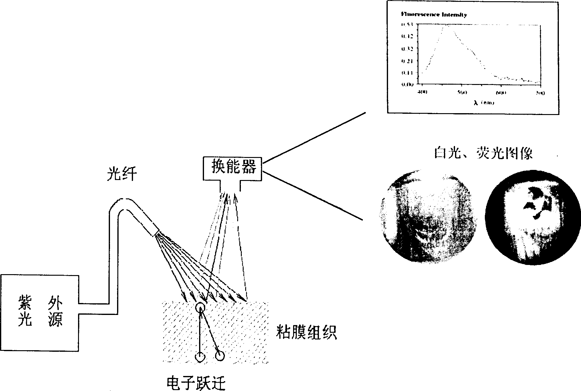
[0022] Based on biochemistry, the present invention applies spectral technology to the spectral detection of human tissue, and invents a "light biopsy" diagnostic instrument (LIF technology: Laser-induced fluorescence) that has been recognized internationally to detect precancerous lesions. The diagnostic methods and diagnostic criteria of LIF technology have been recognized internationally.

[0023] About the principle of LIF technology:

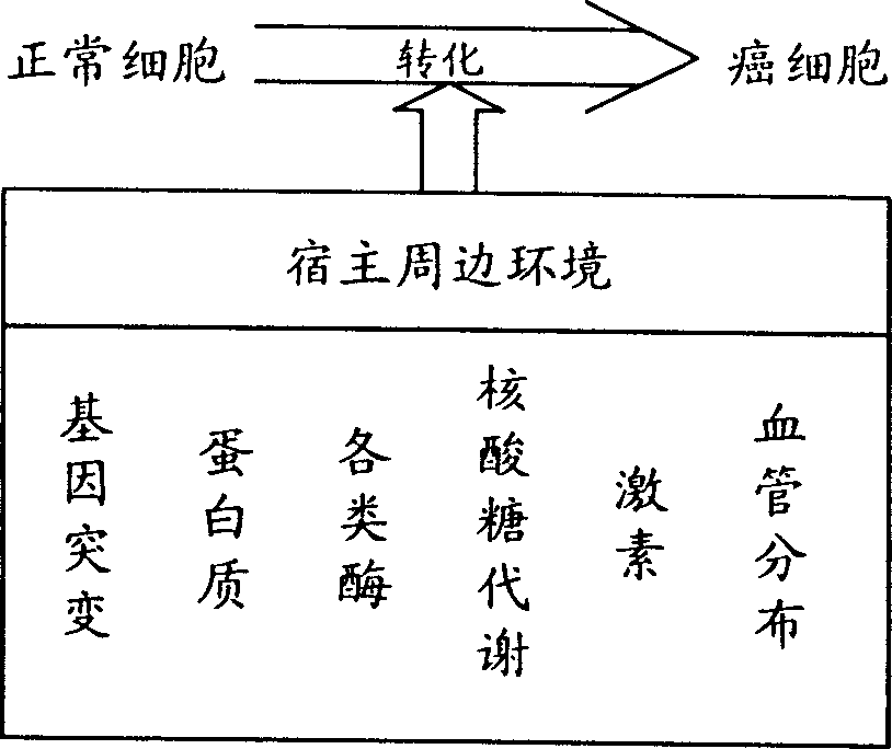
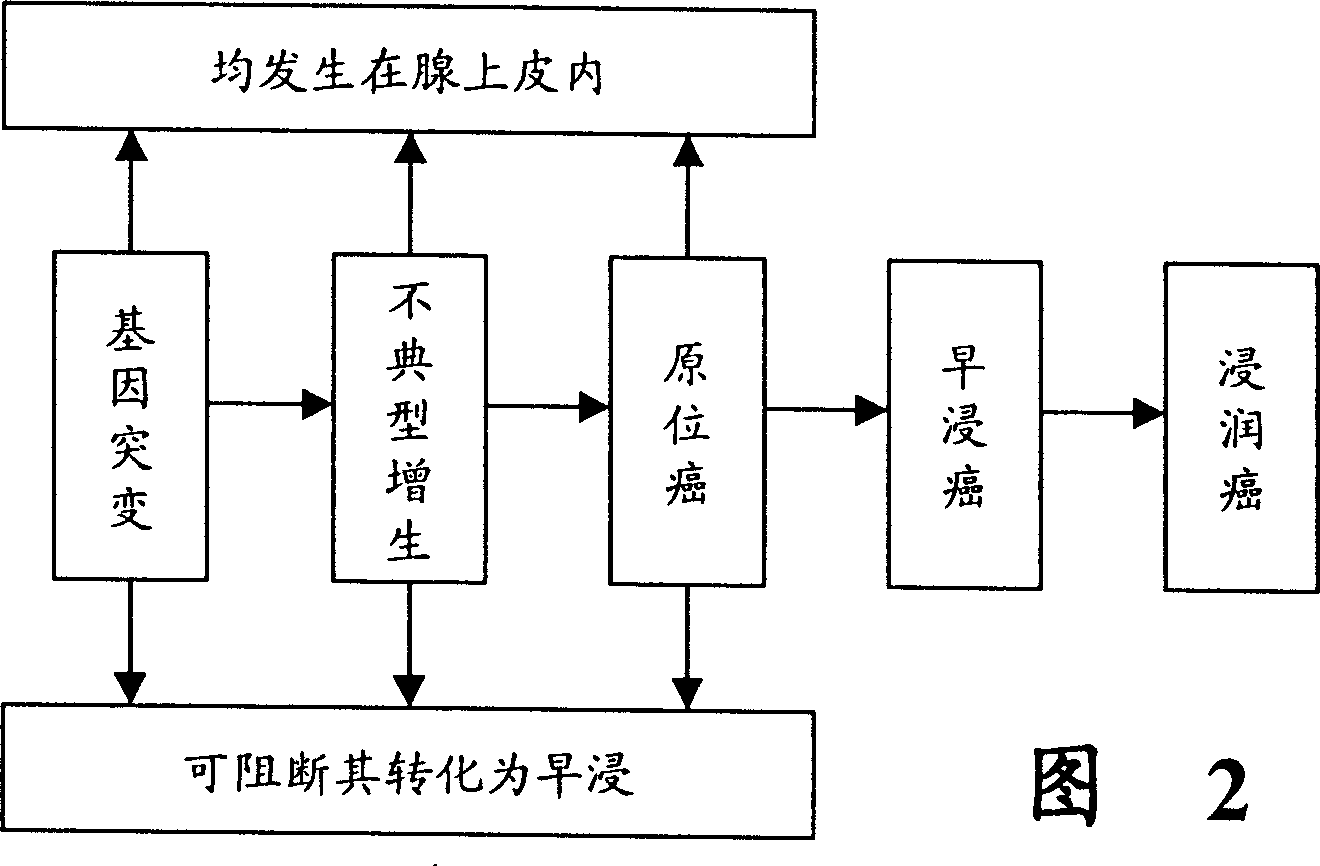
[0024] see first figure 1 , the basic biological characteristics of cancer cells are malignant proliferation, poor differentiation, invasion and metastasis, which are well-known morphological changes. From the point of view of the biochemical changes of normal cells into cancer cells, carcinogenesis begins with gene changes in target cells caused by carcinogenic factors, and then...


PUM


Abstract

An equipment for diagnosing the precancerous lesion with endoscope is composed of light source including cold light source and excited light, optical channel system with optical fibres for transmitting said cold light and excited light, endoscope, and circuit system. Said cold light and excited light can radiate the living tissure to be checked via optical fibres and endoscope. The reflected white light image signals and the inherent fluorescent image signals are received by CCD and then transmitted to circuit system for displaying image. The precancerous lesion can be diagnosed according to image spectrum.

Description

(1) Technical field [0001] The invention relates to a device for diagnosing precancerous lesions, in particular to a medical diagnostic instrument for diagnosing precancerous lesions with an endoscope. (2) Background technology [0002] At present, the reason for the high morbidity and mortality of malignant tumors is that there is no major breakthrough in diagnostics, especially for mucosal tumors, the diagnosis of which still stays in the principles and methods of gross morphological diagnosis, such as doctor's inspection, B-ultrasound, internal medicine, etc. Speculum, CT, MRI, etc. However, this can only be diagnosed as a space-occupying lesion, and it cannot be confirmed whether the space-occupying lesion is a benign tissue or a malignant tissue. Gross morphological diagnostic methods are difficult to distinguish early, moderate and severe dysplasia, because the source of the lesion is subtle biochemical changes in the mucosa, and these subtle changes cannot be detecte...

Claims


Application Information

Patent Timeline
Patent Type & Authority Applications(China)
IPC IPC(8): A61B5/00
CPCA61B5/0071A61B5/0084
Inventor 曾堃虞震芬
Owner SHANGHAI SHENGBIAO SCI & TECH CO LTD
Why Eureka
Try Eureka