Method and equipment for setting medical care device of ultrasound microbubble contrast media to form embolism in capillary vessel

A technology of capillary embolization and ultrasonic microbubbles, which is applied in the direction of ultrasonic therapy, pharmaceutical formulations, preparations for in vivo tests, etc.

Inactive Publication Date: 2004-08-11
JIANGSU HANMEI TECH
View PDF0 Cites 9 Cited by
  • Summary
  • Abstract
  • Description
  • Claims
  • Application Information

AI Technical Summary

Problems solved by technology

Ultrasound-induced microbubble contrast agents lead to intravascular thrombus, especially the formation of capillaries, there is no report at home and abroad

Method used


Image

  • Method and equipment for setting medical care device of ultrasound microbubble contrast media to form embolism in capillary vessel
  • Method and equipment for setting medical care device of ultrasound microbubble contrast media to form embolism in capillary vessel
  • Method and equipment for setting medical care device of ultrasound microbubble contrast media to form embolism in capillary vessel

Examples


Embodiment Construction

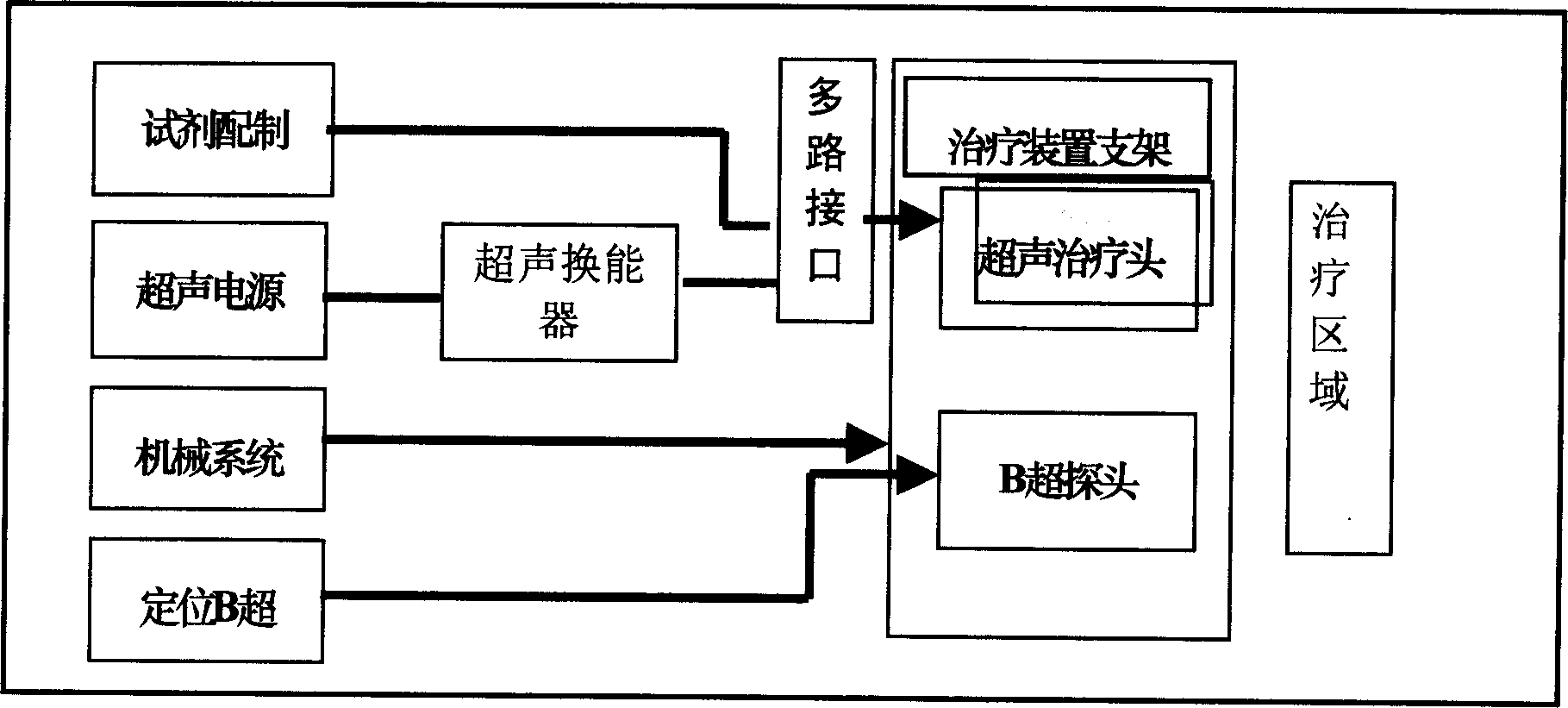
[0024] The present invention uses B-ultrasound positioning (X-ray and CT positioning can also be used) to determine the treatment site and area during operation, and injects the prepared microbubble reagent into the patient's peripheral blood vessels or injects the microbubble reagent into the patient's peripheral blood vessels through an interventional catheter. The position to be treated is then irradiated with low-frequency and low-power ultrasound to the treatment site or area. Ultrasonic microbubble contrast agent is injected as a capillary embolization agent. Under the guidance of CT or B-ultrasound, the area that needs to be embolized is determined, typically such as a tumor area. Ultrasonic energy is transmitted to the area where the agent is injected, and the capillary will form an embolism.

[0025] The device of the present invention is prior art, and its parameter selection is: the ultrasonic wave of effect adopts the ultrasonic wave of low energy and low fr...


PUM


Abstract

An apparatus for using ultrasonic microvesicle contrast medium to generate the thrombus of chosen blood capillary is composed of an ultrasonic microvesicle contrast medium generating injector, a region locating unit for determining the position where thrombus is needed, and an ultrasonic therapeutic unit.

Description

1. Technical field [0001] The invention relates to a setting method and device of a medical device for forming capillary embolism by an ultrasonic microbubble contrast agent. It is especially used for localization and regional formation of capillary embolization for the treatment of tumors and malignant tumors. 2. Background technology [0002] Ultrasound microbubble contrast agent is currently a reagent used in ultrasound detection, mainly used for ultrasound myocardial imaging, see "The Effect of Ultrasound Irradiation on the Stability of Acoustic Vibration Microbubbles", Zha Daogang et al., Journal of First Military Medical University, 1999 Issue 5, Volume 19, contrast agent microbubble concentration and size are the most important factors affecting myocardial acoustic imaging. The study reported that 2×2×4 factorial analysis method was used to analyze the influence of different ultrasonic irradiation conditions on the concentration and diameter of contrast agent microbu...

Claims


Application Information

Patent Timeline
Patent Type & Authority Applications(China)
IPC IPC(8): A61B18/00A61K49/00A61N7/00
Inventor 吴巍姜藻
Owner JIANGSU HANMEI TECH
Why Eureka
Try Eureka