Image display apparatus, image display method, and image display program

An image display device and image display technology, which can be applied to medical images, image enhancement, image analysis, etc., and can solve the problem of a large number of images

Inactive Publication Date: 2006-05-24
OLYMPUS CORP
View PDF1 Cites 15 Cited by
  • Summary
  • Abstract
  • Description
  • Claims
  • Application Information

AI Technical Summary

Problems solved by technology

[0005] However, in the above-mentioned capsule endoscope, unlike ordinary endoscopes, since each organ is photographed after being swallowed by the examinee until it

Method used


Image

  • Image display apparatus, image display method, and image display program
  • Image display apparatus, image display method, and image display program
  • Image display apparatus, image display method, and image display program

Examples


Embodiment Construction

[0033] Hereinafter, the best mode for carrying out the present invention will be described in detail with reference to the accompanying drawings.

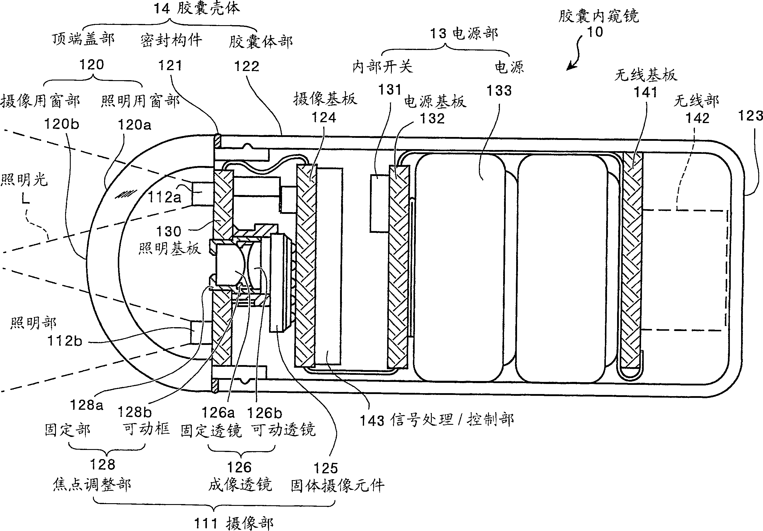
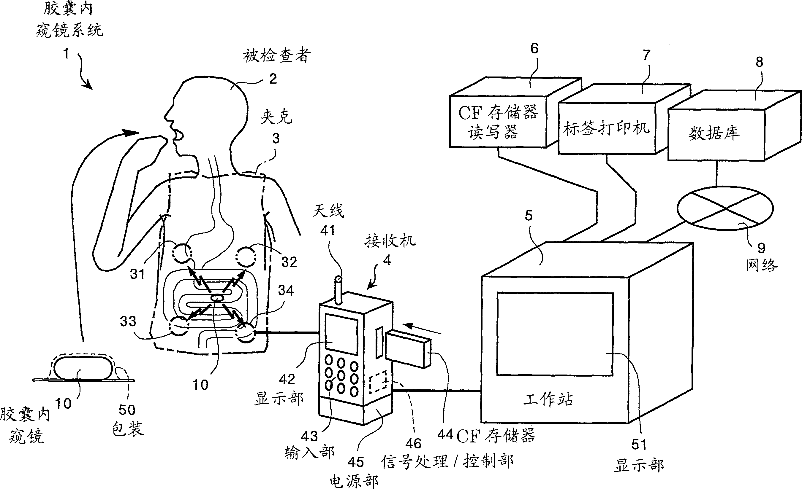
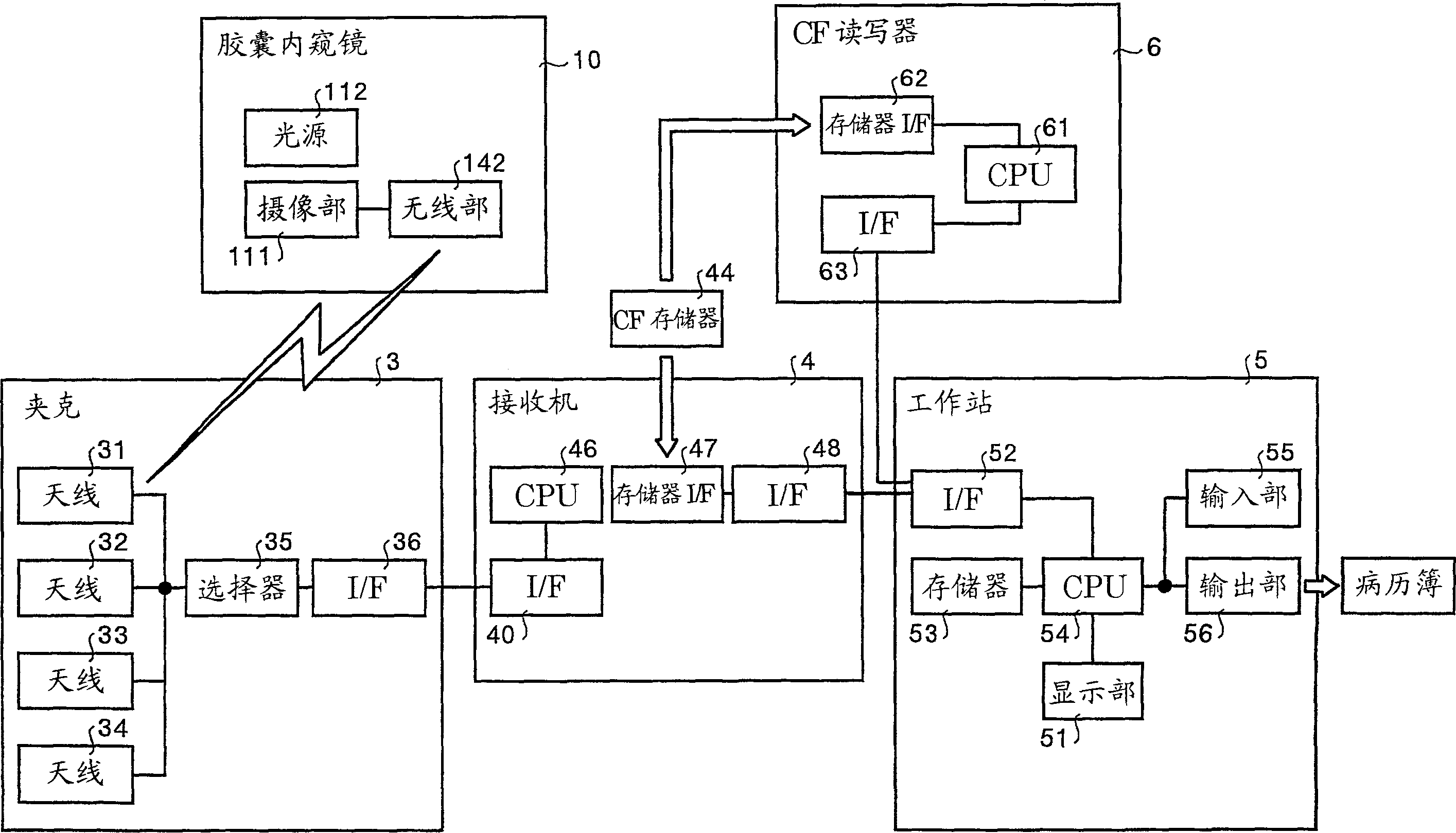
[0034] First, for the capsule endoscope used in one embodiment of the present invention, refer to figure 1 Describe the overall structure. figure 1 It is a schematic diagram showing the internal structure of the capsule endoscope according to this embodiment. Such as figure 1 As shown, the capsule endoscope 10 is composed of the following parts: an imaging unit 111 capable of capturing images inside the body cavity, illuminating units 112a, 112b illuminating the inside of the body cavity, a power supply unit 13 for supplying power to them, and at least the imaging unit described above. part 111, the above-mentioned lighting part 112 and the capsule casing 14 of the above-mentioned power supply part 13.

[0035] Here, the capsule casing 14 related to the present embodiment is composed of a top end cover part 120 and a capsule bod...


PUM


Abstract

An average color bar (507) representing the entire imaging period of images captured by the capsule endoscope in time series is displayed. The checked images in all captured images are displayed in a list in the checked image display column (504B), and it is calculated which time in the imaging period each checked image corresponds to, and the scale of the average color bar (507) is used to set the average color bar (507). (507) mark and display with numbers corresponding to each inspection image. As a result, the searchability of the image obtained by shooting the inside of the body is improved, and it is possible to easily identify which organ the displayed image is.

Description

technical field [0001] The present invention relates to, for example, an image display device, an image display method, and an image display program. Background technique [0002] In recent years, among endoscopes, swallowable capsule endoscopes have appeared. In this capsule endoscope, an imaging function and a wireless function are provided. Capsule endoscope is a structure that, for observation (inspection), successively photographs organs such as the stomach and small intestine during the observation period after being swallowed by the patient's mouth and being discharged naturally from the human body (see Japanese Patent Application Laid-Open No. 11-225996 Bulletin). [0003] During this observation period, image data taken inside the body by the capsule endoscope is sequentially transmitted to the outside by wireless communication and stored in a memory. By carrying the receiver with the wireless communication function and memory function, the patient can move freel...

Claims


Application Information

Patent Timeline
IPC IPC(8): A61B1/04G02B23/24A61B1/00A61B1/05H04L1/00H04N5/225
CPCA61B1/00009A61B1/00016A61B1/00022A61B1/0005A61B1/041A61B5/073A61B5/6805G06T2207/10016G06T2207/10068G06T2207/30028H04L1/004G06T7/90G16H40/63G16H30/40A61B1/0004A61B1/04
Inventor 本多武道药袋哲夫
Owner OLYMPUS CORP
Why Eureka
Try Eureka