Systems, methods and apparatus for dual mammography image detection

A mammography imaging, breast technology, applied in the direction of equipment for radiological diagnosis, mammography, ultrasound/sonic/infrasound equipment testing/calibration, etc.

Active Publication Date: 2006-12-06
GENERAL ELECTRIC CO
View PDF0 Cites 15 Cited by
  • Summary
  • Abstract
  • Description
  • Claims
  • Application Information

AI Technical Summary

Problems solved by technology

Much of the hardware, such as computers and monitors, is duplicated, though, because the systems are built and sold separately

Method used


Image

  • Systems, methods and apparatus for dual mammography image detection
  • Systems, methods and apparatus for dual mammography image detection
  • Systems, methods and apparatus for dual mammography image detection

Examples


Embodiment Construction

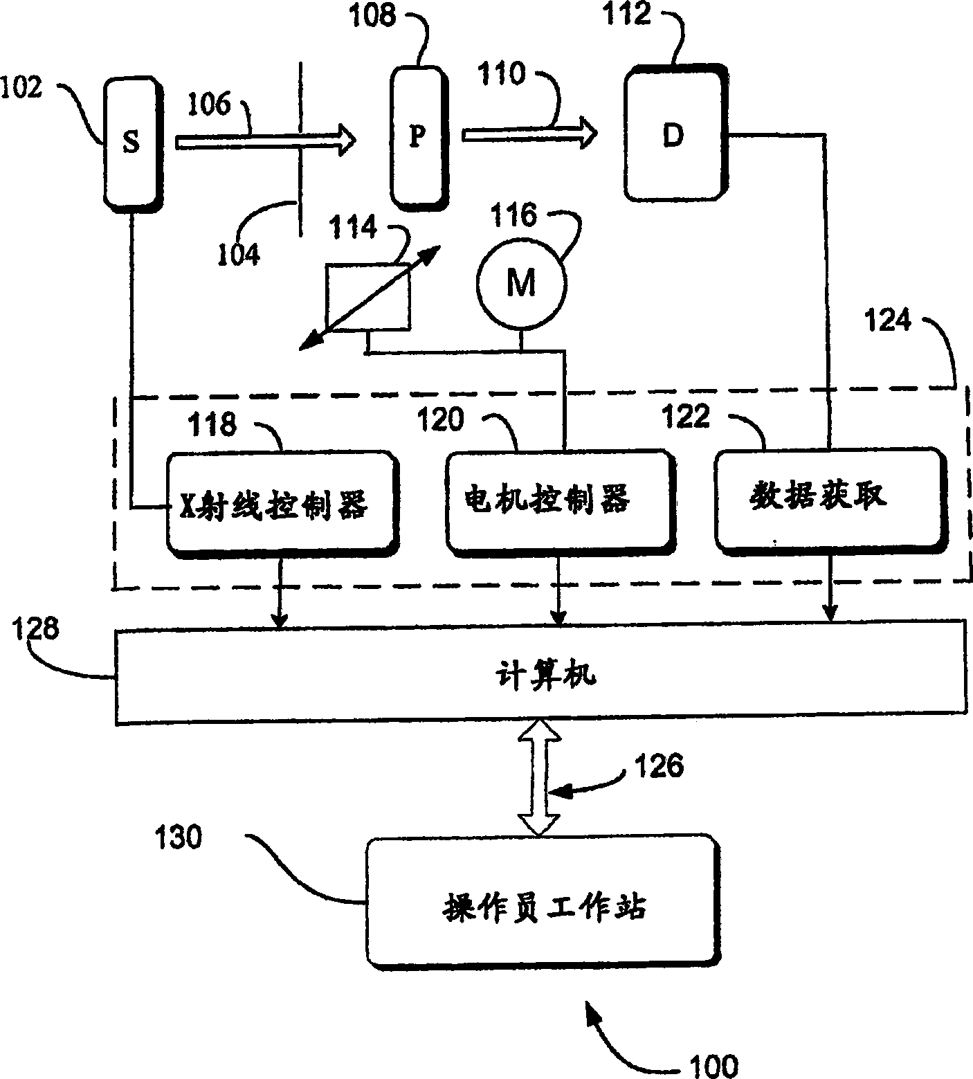
[0026] In the following detailed description, reference is made to the accompanying drawings which form a part of the specification and which are given by way of illustrative embodiments that can be practiced. These embodiments are described in sufficient detail to enable those skilled in the art to practice these embodiments, and it is to be understood that other embodiments may be employed and may be made without exceeding the scope of these embodiments Logical, mechanical, electronic and other changes. Therefore, the following detailed description is not given in a limiting sense.

[0027] This detailed description is divided into four sections. In the first part, a system-level overview is presented. In the second part, the method of implementation is introduced. In the third section, the hardware and operating environment in which these embodiments are practiced is described. In the fourth section, special implementations are introduced. Finally, in the fifth section...


PUM


Abstract

Systems and methods are provided by which a mammography imaging system offers X-ray and ultrasound imaging that allows sharing of common hardware such as the computer and display. Small regions of interest are imaged with X-ray at higher image quality by using a second sensor with higher DQE than the full-field sensor can obtain. In some embodiments a specialized chamber is provided for securing the anatomy to a fixed location, ultrasound image data is collected along with ultrasound probe location and orientation data from sensors on a handheld probe from which data images can be viewed directly, or used to reconstruct tomographic images of any desired cross-section, or used for various ' 3 -D' image visualization methods. An imaging schedule defined by location and orientation of an ultrasound probe is used to generate a three-dimensional ultrasound image.

Description

technical field [0001] The present invention relates generally to mammography imaging systems and, more particularly, to images with higher detectable quantum efficiency. Background technique [0002] The use of X-ray technology to provide two-dimensional images of breast tissue for the purpose of diagnosing tumors or other abnormalities is being widely used. However, X-ray imaging of breast tissue has inherent limitations, and mammograms can only provide planar images of three-dimensional objects. [0003] The detectable quantum efficiency ("DQE") of an image is a traditional measure of X-ray image quality. Simply put, DQE is the resolution of the detector. For a given detector and dose technique, the DQE is constant over the entire image. [0004] When a possible area of ​​medical interest is marked on a mammogram, the bulge or depth of the object area in the two-dimensional image of the breast may be indeterminate. Existing digital X-ray imagers can give full-view or ...

Claims


Application Information

Patent Timeline
Patent Type & Authority Applications(China)
IPC IPC(8): A61B8/13A61B8/00A61B6/03
CPCA61B6/548A61B8/4209A61B8/0825A61B8/4218A61B6/04A61B8/483A61B6/502A61B6/4266A61B8/565A61B8/58
Inventor D·C·诺伊曼H·瓦菲
Owner GENERAL ELECTRIC CO
Why Eureka
Try Eureka