Method and apparatus for non-compressed evaluation of tissue specimens

a tissue specimen and non-compressed technology, applied in the field of tissue specimen non-compressed evaluation, can solve the problems of loss of relative orientation of the sample in the transition, the surgeon's inability to remove the appropriate amount of tissue from the patient, etc., and achieve the effect of ensuring the relative position and angle of the tissue sample, and facilitating the transport of the sampl

Inactive Publication Date: 2018-07-10
RUPLEY DANIEL
View PDF11 Cites 0 Cited by
  • Summary
  • Abstract
  • Description
  • Claims
  • Application Information

AI Technical Summary

Benefits of technology

[0018]The present invention achieves the above objectives by providing apparatus primarily composed of three elements: an end cap, a top cap, and a flexible, transparent piece of material. The tissue, upon removal from the patient, will feature a physical stitch in the tissue sample oriented at one of the six orthogonal positions of the patient's body. The handler of the tissue sample will place the sample on the piece of flexible, transparent material, and orient the stitch of the tissue sample with a notch provided on the flexible, transparent material. The flexible, transparent material also features a slit or series of slits in its side, so that the handler may then gently roll the flexible material around the tissue sample and lock it into a spherical configuration via the slit or slits on the side of the material. Given the variety of sizes of material and slit positions available in the present invention, the handler would be able to roll the sheet of material around the tissue sample in such a manner that the tissue sample will be suspended within the material, but not compressed or unduly distorted. After the sample is encased in the transparent material, the rolled material is inserted into the end cap of the apparatus. This end cap features a series of lined, circular grooves, meant to receive the rolled material. The end cap, crucially, also features an indicator conveying the appropriate location of the stitch from the tissue sample. When the tissue sample, encased in the rolled material, is placed in this appropriate position of the end cap, the integrity of the relative positions and angles of the tissue sample are ensured. The top cap is then placed on the opposite end of the tissue sample, securing the placement of the flexible, transparent material. By capping both ends, the handler may easily transport the sample to the appropriate evaluator, such as the pathologist, for analysis. In such transport, the relative position of the cancerous cells within the tissue sample will always remain certain, given the orientation of the stitch in the sample. Because the sample is encased in a transparent tube, each orthogonal angle of the sample, and its corresponding margins, may be evaluated, and such information may then be conveyed to the surgeon while on-site and available to most appropriately conclude or continue the surgical procedure.

Problems solved by technology

When a patient undergoes a surgery to remove a cancerous lump, one of the challenges a surgeon faces is removal of the appropriate amount of tissue from the patient.
However, when a sample is suspended in a manner such that it may be evaluated from every angle, the relative orientation of the sample may be lost in the transition.
However, as outlined below, the prior art has not provided for a container providing a non-compressed, orthogonal view of the tissue sample, which would fully allow the viewer or radiographic instrument to evaluate whether every margin of the removed sample is free from cancerous cells, while at the same time providing a quick and intuitive method of orienting the sample inside the container, which would allow for on-site evaluation of the tissue sample, thereby allowing a surgeon to remove the appropriate amount of tissue from the patient without requiring additional surgery following an off-site evaluation of the sample.

Method used


Image

  • Method and apparatus for non-compressed evaluation of tissue specimens
  • Method and apparatus for non-compressed evaluation of tissue specimens
  • Method and apparatus for non-compressed evaluation of tissue specimens

Examples


Embodiment Construction

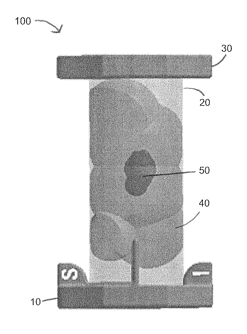
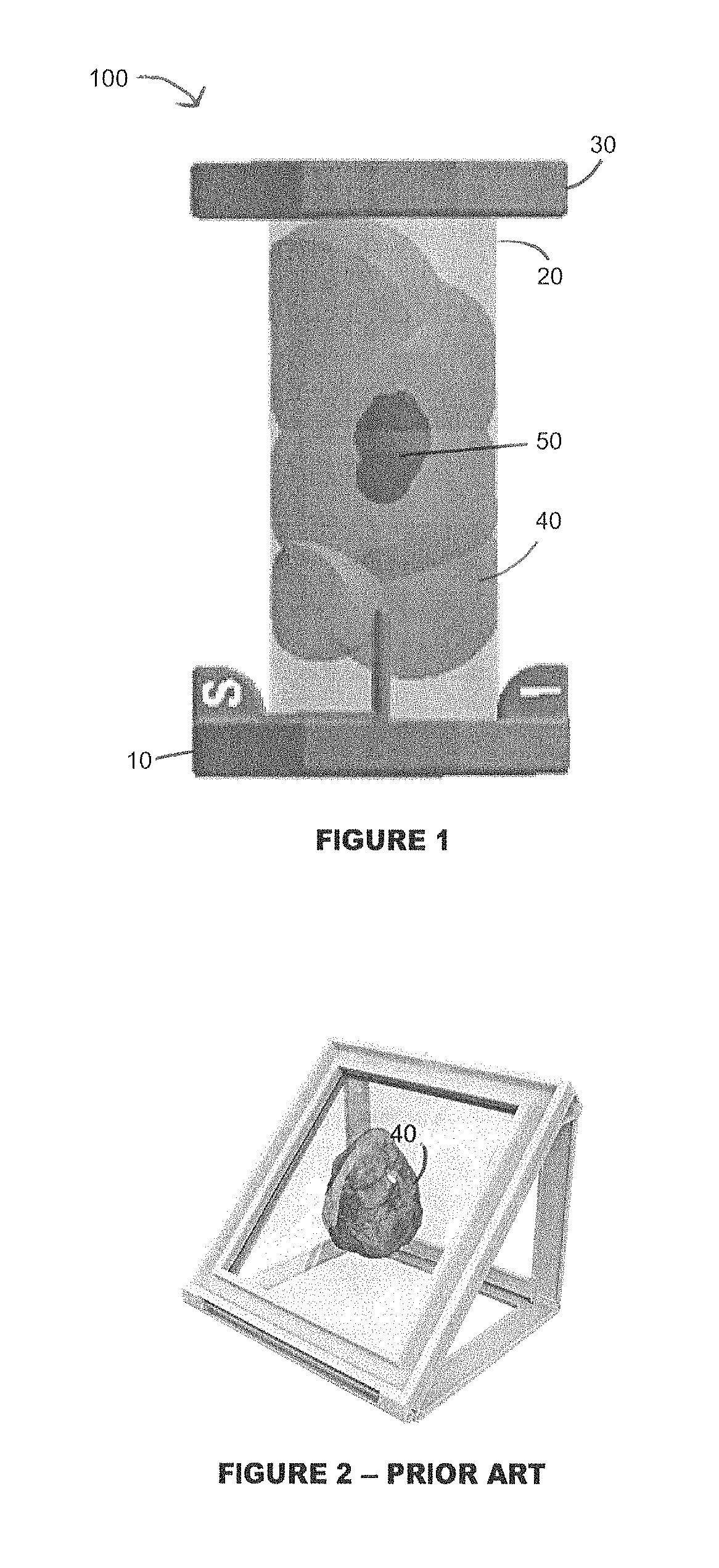
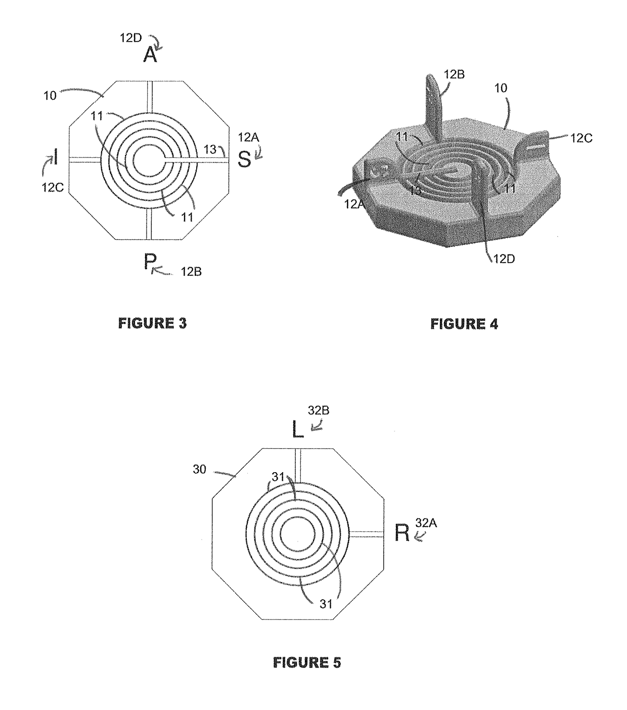
[0031]Referring to FIG. 1 generally, the present invention provides for an apparatus 100 featuring an end cap 10, a flexible plastic sheet 20, and top cap 30. The end cap 10 features a series of lined grooves embedded in the end cap. The flexible plastic sheet 20, when rolled into a round or spherical configuration, may be fitted into one of lined grooves embedded in the end cap 10. After being placed in the appropriate circular groove, the top cap 30, which also features a series of lined grooves embedded into one side of the top cap 30, is placed on the top end of the rolled plastic sheet 20 and the plastic sheet 20 fit into the appropriate lined groove of the top cap 30. Upon placement of the top cap 30 and end cap 10 on the rolled plastic sheet 20, the tissue specimen 40, which may include cancerous cells 50, contained within the sheet 20 will remain suspended and ready for examination by the handler of the apparatus 10.

[0032]FIG. 2 is an example of the conventional manner of en...


PUM

PropertyMeasurementUnit
widthaaaaaaaaaa
heightaaaaaaaaaa
diameteraaaaaaaaaa

Abstract

The present invention provides a method and apparatus for evaluating the margins of a surgically-removed tissue specimen, such as a breast tissue specimen, to determine whether sufficient fatty tissue has been removed from around the lesion or cancerous point. The instant invention provides a solution to the problem of on-site evaluation of the margin sufficiency during the surgical procedure, in that it provides surgeons with an orthogonal view of all sides of the tissue specimen to be evaluated. The specimen evaluation device provides for properly-oriented examination of the removed specimen in a non-compressed, undistorted manner, both by visual inspection and through radiographic evaluation. Through this examination of the properly-oriented specimen, the surgeon may quickly and more accurately be informed of whether there remain cancerous cells in the margins surrounding the sample, which are meant to be free of cancerous cells. Upon evaluation of the margins of the removed sample, the surgeon may then make an on-site determination on whether to proceed with additional surgery or complete the surgical procedure.

Description

BACKGROUND OF THE INVENTION[0001]The present invention provides a method and apparatus for evaluating the margins of a surgically-removed tissue specimen, such as a breast tissue specimen, to determine whether sufficient fatty tissue has been removed from around the lesion or cancerous point. The instant invention provides a solution to the problem of on-site evaluation of the margin sufficiency during the surgical procedure, in that it provides surgeons with an orthogonal view of all sides of the tissue specimen to be evaluated. The specimen evaluation device provides for properly-oriented examination of the removed specimen in a non-compressed, undistorted manner, both by visual inspection and through radiographic evaluation. Through this examination of the properly-oriented specimen, the surgeon may quickly and more accurately be informed of whether there remain cancerous cells in the margins surrounding the sample, which are meant to be free of cancerous cells. Upon evaluation o...

Claims


Application Information

Patent Timeline
Patent Type & Authority Patents(United States)
IPC IPC(8): G01N3/00B01L3/00G01N1/36
CPCB01L3/505G01N1/36B01L2300/0832B01L2300/042B01L2200/025A61B10/0041A61B10/0096B01L2200/023
Inventor RUPLEY, DANIEL
Owner RUPLEY DANIEL
Why Eureka
Try Eureka