Method and system for multi-shot spiral magnetic resonance elastography pulse sequence

a magnetic resonance elastography and pulse sequence technology, applied in the field of multi-shot spiral magnetic resonance elastography pulse sequence, can solve the problems of not being able to achieve the quality of mre property distribution, not all types of mr techniques are suitable for every purpose, and not being able to accurately achieve the effect of mr property distribution,

Active Publication Date: 2019-04-16
THE BOARD OF TRUSTEES OF THE UNIV OF ILLINOIS
View PDF26 Cites 3 Cited by
  • Summary
  • Abstract
  • Description
  • Claims
  • Application Information

AI Technical Summary

Problems solved by technology

Mechanical palpation of the tissue can be utilized, but is not an accurate technique and can be invasive depending on the tissue that is being analyzed.
However, not all types of MR technique are suitable for every purpose.
Model-data mismatch can also limit the achievable quality of MRE property distributions, as any continuum tissue model is only an approximation of the net effect of enormous numbers of complicated micro-scale interactions.
Contemporary processes utilize single-shot, spin-echo echo-planar imaging (EPI) that can provide rapid acquisition, but in high-resolution protocols, single-shot EPI sequences suffer from very long readout durations.
Long readouts result in large distortions from field inhomogeneity and also require long echo times, thus reducing SNR and ultimately limiting the resolution achievable in a short scan time.
Single-shot sequences suffer from very long readout times in high-resolution acquisitions.
The long readout time for the single-shot sequences can lead to significant distortions in the presence of magnetic field inhomogeneities and susceptibility differences at air-tissue interfaces.
These distortions not only degrade the quality of the image, but may also lead to model / data mismatch during the inversion, which produces errors in the resulting estimates of mechanical properties.
As an example, geometric distortions from field inhomogeneity can cause compression and stretching of spatial information in certain regions, which can impact the inversion algorithms for determining mechanical properties.
Multi-shot imaging in the presence of motion-encoding gradients can include phase errors between shots, such as from bulk motion not related to the vibrations of interest.
This phase error can lead to phase cancellation and signal loss.
For an MRE multi-shot sequence, a likely source of phase error arises from variations in mechanical actuation, which may be strained when loaded, such as with the weight of the human head.
Additionally, phase errors may result from brain pulsation since the sequence is not cardiac gated, and also small subject motions not controlled in the experiment.
The amplitude of RBM more closely reflects the applied displacement from the actuator, and thus, any variations can lead to phase errors between shots.
In MRE, these phase errors can be attributed to variations in the amplitude of motion imparted by the actuator, which effectively contributed an additional, erroneous RBM phase component to the signal.
Signal loss in the MR images leads to an increase in noise in the displacement fields calculated from the phase, and significantly decreases the OSS-SNR to levels that are below the threshold required for accurate inversion, which is approximately 3.0.
Additionally, anatomical features visible in the corrected data are obscured in the uncorrected data.
Additionally, the corticospinal tracts inferior to the corona radiata appear as very stiff regions to the left and right of the ventricles in the axial view, though are not uniform bilaterally.
Though the phantom study data of Example 1 did not exhibit significant motion-induced phase errors, such errors were prevalent in the in vivo brain data from Example 1 due to the increased loading of the head on the actuator.

Method used


Image

  • Method and system for multi-shot spiral magnetic resonance elastography pulse sequence
  • Method and system for multi-shot spiral magnetic resonance elastography pulse sequence
  • Method and system for multi-shot spiral magnetic resonance elastography pulse sequence

Examples


example 1

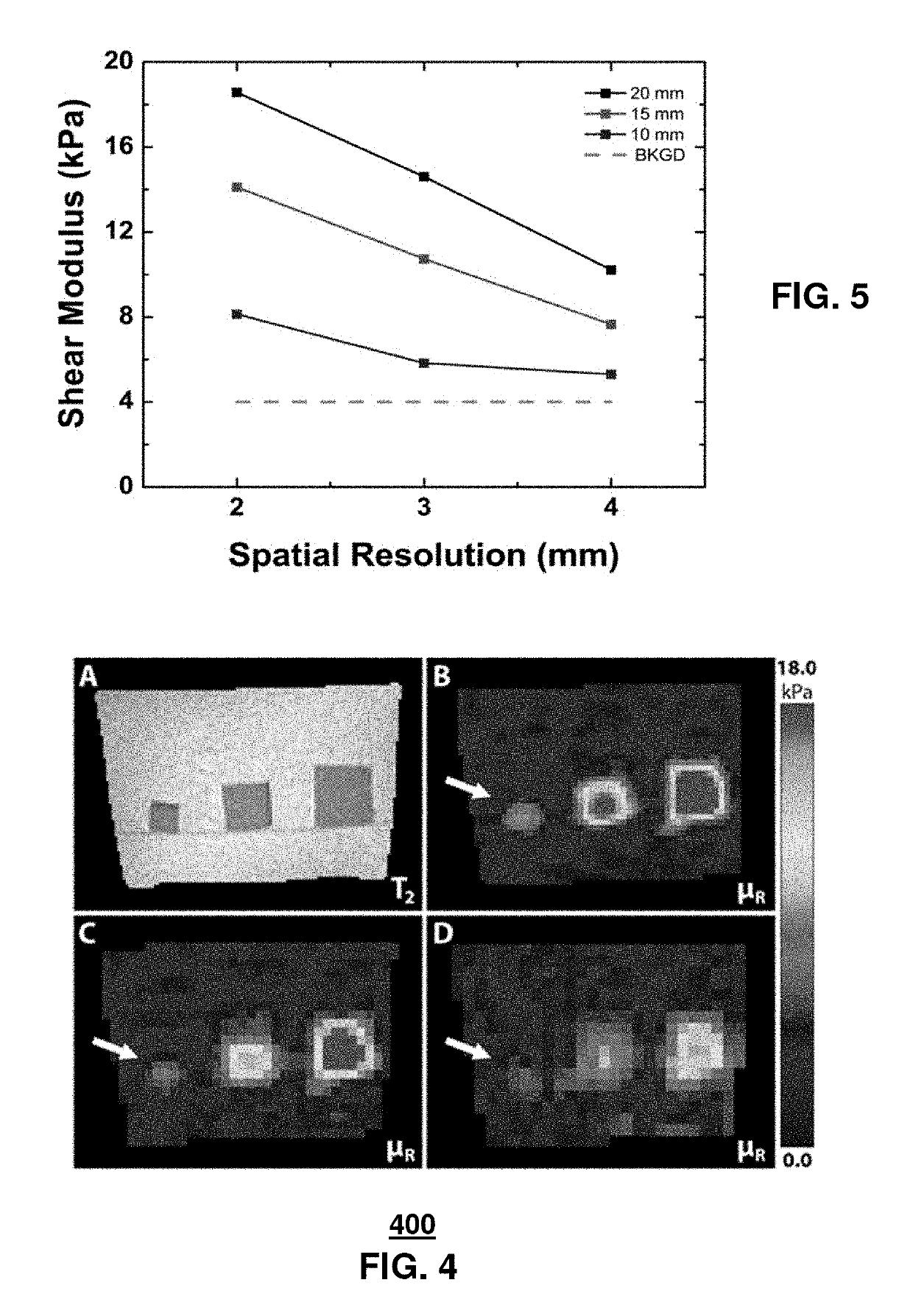
[0049]The phantom used in the experiments was a rectangular parallelepiped composed of agarose gel (1%) with three stiffer inclusions (2%) embedded. The inclusions were cubes of three different sizes: 10, 15, and 20 mm on a side. Shear waves were generated by vibrating the lower surface of the phantom at 100 Hz.

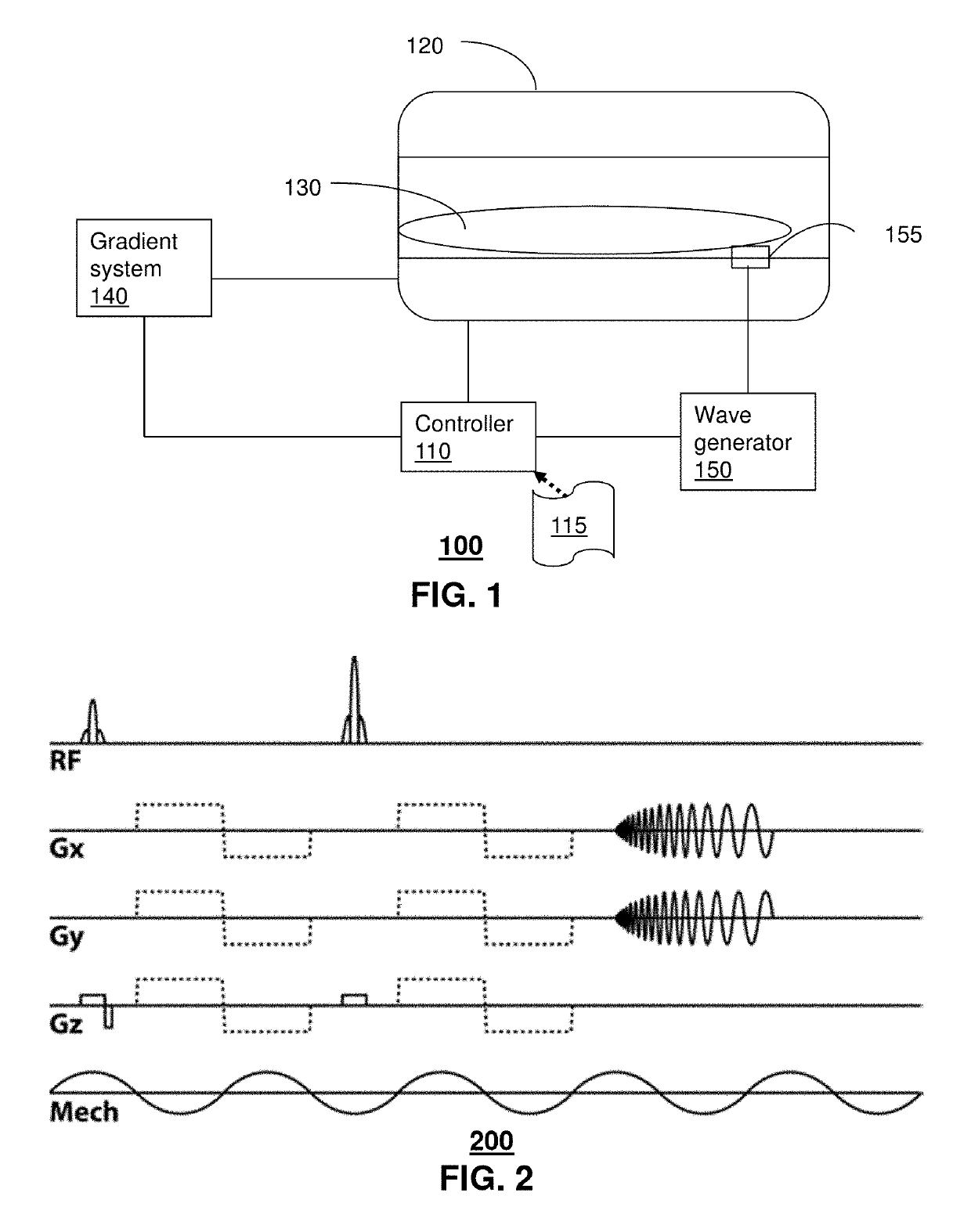
[0050]Images were acquired using a multi-shot spiral MRE sequence with bipolar motion-encoding gradients matched in period to the vibration frequency of 100 Hz, as described above with respect to system 100. MRE encoding was performed on each of the three cardinal gradient directions independently, and a single period of vibration was sampled with eight evenly spaced points. Six interleaved spiral shots were used to cover k-space with a 64×64 matrix size for the phantom, with resolution achieved by adjusting the overall field-of-view: 128, 192, and 256 mm Twenty slices were acquired with 2, 3, or 4 mm thickness, corresponding to the in-plane resolution resulting in isotropic ...

example 2

[0054]In vivo brain tissue examination was performed utilizing an exemplary embodiment of the present disclosure. Actuation during the brain MRE study was performed with a system that comprised a remote electromagnetic shaker having a long rod that vibrates a custom cradle the subject's head rests on, similar to the one used in other brain MRE studies. (See Sack et al. Non-Invasive Measurement of Brain Viscoelasticity Using Magnetic Resonance Elastography. NMR Biomed 2008; 21:265-271, the disclosure of which is hereby incorporated by reference). The actuator imparted a nodding motion to the head at the driving frequency of 50 Hz. Imaging was performed in the same manner as the phantom study with motion encoding along three axes and eight samples over a single period. Imaging parameters included: six k-space interleaves; 256 mm field-of-view; 128×128 matrix; 20 axial slices (2 mm thick) in the region of the corpus callosum; 2000 / 55 ms repetition / echo times. This acquisition resulted ...


PUM


Abstract

Aspects of the subject disclosure include a system that applies magnetic resonance elastography to a sample to obtain uncorrected k-space data where the magnetic resonance elastography utilizes a multi-shot spin-echo sequence with variable density spiral readout gradients, and adjusts the uncorrected k-space data to corrected k-space data by adjusting a k-space trajectory by shifting a center point for each shot to a new center point according to signal intensity and by adjusting a phase for each shot based on a phase offset that is determined according to the signal intensity.

Description

PRIOR APPLICATION[0001]The present application claims the benefit of priority to U.S. Provisional Application No. 61 / 765,919 filed on Feb. 18, 2013, the disclosure of which is hereby incorporated herein by reference in its entirety.FIELD OF THE DISCLOSURE[0002]The subject disclosure relates to a method and system for multi-shot spiral magnetic resonance elastography pulse sequence.BACKGROUND[0003]Identifying mechanical properties of tissues (e.g., brain, liver, breast and so forth) or other samples can be useful for a number of purposes including diagnosis. Mechanical palpation of the tissue can be utilized, but is not an accurate technique and can be invasive depending on the tissue that is being analyzed.[0004]Magnetic Resonance (MR) techniques are being employed for various imaging purposes. However, not all types of MR technique are suitable for every purpose. The type of MR technique and / or the type of sample being analyzed can affect the quality of the images, as well as other...

Claims


Application Information

Patent Timeline
Patent Type & Authority Patents(United States)
IPC IPC(8): G01V3/00G01R33/563G01R33/565G01R33/48
CPCG01R33/56358G01R33/56509G01R33/4824
Inventor SUTTON, BRADLEYGEORGIADIS, JOHN G.JOHNSON, CURTIS
Owner THE BOARD OF TRUSTEES OF THE UNIV OF ILLINOIS
Why Eureka
Try Eureka