Biological heart valve replacement, particularly for pediatric patients, and manufacturing method

a heart valve and biotechnology, applied in the field of biological heart valve replacement, can solve the problems of lack of growth-adaptive capacity, prone to thromboembolic complications of metal-based “mechanoprostheses” and other problems, to achieve the effect of supporting the maintenance of a physiological geometry of the heart valve and improving the “growth” adaptation

Active Publication Date: 2019-05-21
UNIV ZURICH
View PDF8 Cites 0 Cited by
  • Summary
  • Abstract
  • Description
  • Claims
  • Application Information

AI Technical Summary

Benefits of technology

[0019]The present invention overcomes the disadvantages of currently known biological heart valve replacements by having integrated tubular growth zones, which provide the capacity of gradual radial expansion according to the physiological growth of children. The invention thus combines the advantages of clinically used biological (homologous or xenogenic) prostheses, which are not plagued by surface thrombogenicity but do not have growth-adaptive properties, and of certain non-biological heart valve prostheses designed to allow size adaptation but having the disadvantage of substantial thrombogenicity.
[0036]A further advantage of the proposed growth zones could be that they may serve as entrance point for autologous host cells repopulating the tissues and could ultimately also improve the recellularization of the non-viable bioprosthetic implant tissues.

Problems solved by technology

The metal-based “mechanoprostheses” are prone to thromboembolic complications and lack growth-adaptive capacities (Schoen, 2008).
While bioprosthetic materials, e.g. fabricated from glutaraldehyde-“fixed” xenogenic or homogenic native tissues, are not associated with an increased risk of clotting activation, they are still limited by the lack of growth-adaptive capacities of these implants.
This implies that these young patients currently have to undergo repeated reoperations causing increased morbidity and mortality (Talwar et al., 2012).
However, while for tissue engineered arteries adequate function (and growth adaptation) could be demonstrated and the technology has also entered first-in-man clinical trials in Japan (Hibino et al., 2010) and the US (Vogel et al., 2011; Dolgin et al., 2011), the development of tissue engineered heart valves is experiencing obstacles partly derived from the complexity of the physiological environment of heart valves (Weber et al., 2011; Schmidt et al., 2010).
Therefore, so far no tissue engineered heart valve has entered routine clinical practice, and therapy for patients with heart valve disease remains to be highly limited.

Method used


Image

  • Biological heart valve replacement, particularly for pediatric patients, and manufacturing method

Examples


Embodiment Construction

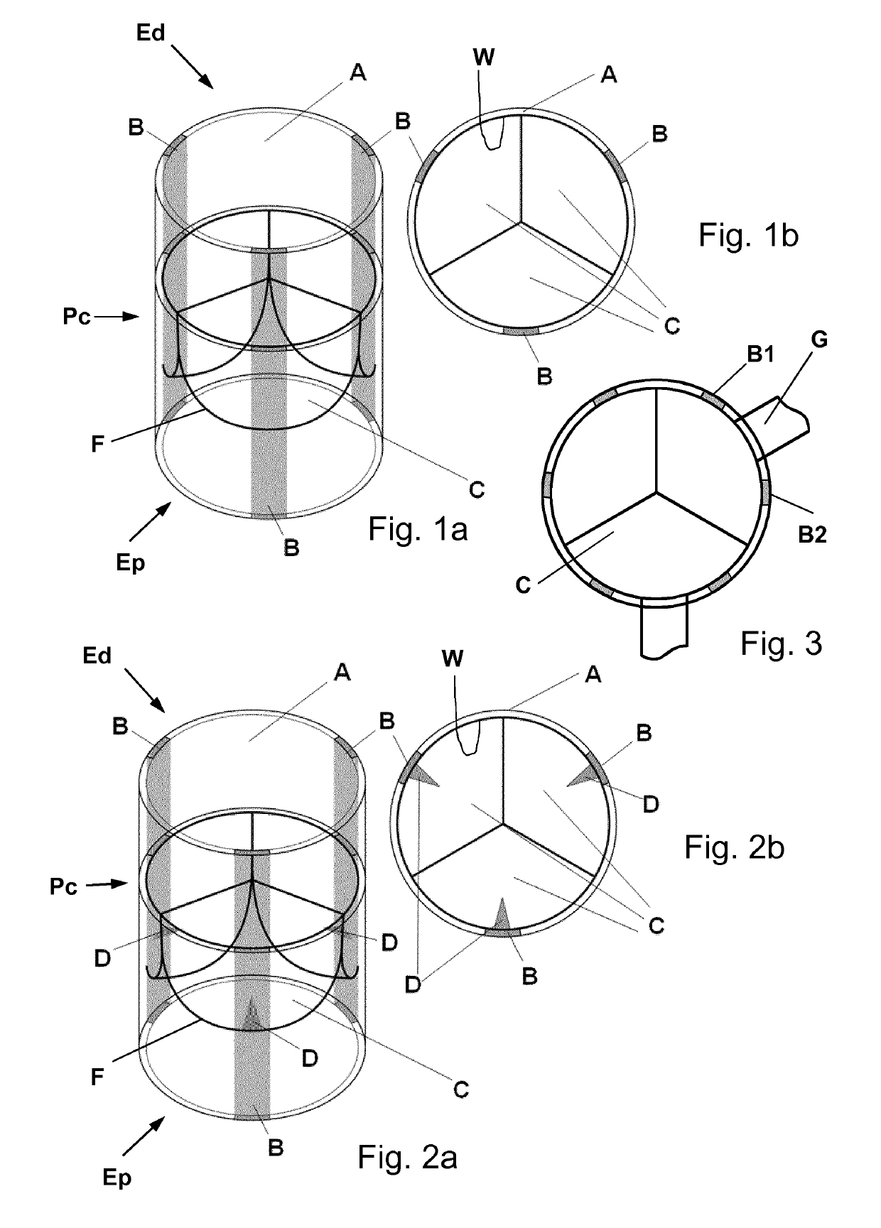
[0050]In the following, exemplary heart valve replacements are illustrated as a three-leaflet or tricuspid valves. However, it will be appreciated by the skilled person that such valve replacements may be configured to have just two leaflets or a larger number of leaflets depending on the intended application.

[0051]The biological heart valve replacement shown in FIGS. 1a and 1b comprises a tubular segment A having a proximal end Ep, a distal end Ed and a central portion Pc arranged between said proximal and distal ends and defining a longitudinal direction of the valve. The valve further comprises three inner leaflets C attached in hinge-like manner to respective connection zones F at an inner wall W region of the central portion. The inner leaflets are movable between a closing position as shown in FIGS. 1a and 1b and an opening position (not shown here) of the valve where the inner leaflets are flipped towards the valve's inner wall. The tubular segment comprises three tubular gro...


PUM

PropertyMeasurementUnit
diameteraaaaaaaaaa
lengthaaaaaaaaaa
lengthaaaaaaaaaa

Abstract

A biological heart valve replacement, particularly for pediatric patients, comprises a tubular segment (A) comprising a proximal end (Ep), a distal end (Ed) and a central portion (Pc) arranged between said proximal and distal ends and defining a longitudinal direction of the valve. The valve further comprises at least one inner leaflet (C) attached in hinge-like manner to a connection zone (F) at an inner wall (W) region of said central portion, each one of said inner leaflets being movable between a closing position and an opening position of the valve. In order to provide growth adaptability, the tubular segment comprises at least one tubular growth zone (B; B1,B2) configured as a longitudinal strip made of a growth-adaptive biomaterial, with the remainder of the tubular segment being made of a non-growth-adaptive biomaterial.

Description

CROSS-REFERENCE TO RELATED APPLICATIONS[0001]This is the U.S. national stage of International application PCT / EP2014 / 070355, filed Sep. 24, 2014 designating the United States and claiming priority to European application EP 13186027.2, filed Sep. 25, 2013.FIELD OF THE INVENTION[0002]The present invention generally relates to a biological heart valve replacement, particularly for pediatric patients. Moreover, the invention relates to a method of manufacturing a biological heart valve replacement, particularly for pediatric patients.BACKGROUND OF THE INVENTION[0003]Heart valve disease represents a major cardiovascular disease worldwide. Besides acquired heart disease, also congenital heart disease (affecting 1% of all life births) is responsible for a major disease global load. Currently used heart valve replacement constructs are fabricated from either metallic or fixed “biological” materials. The metal-based “mechanoprostheses” are prone to thromboembolic complications and lack grow...

Claims


Application Information

Patent Timeline
Patent Type & Authority Patents(United States)
IPC IPC(8): A61F2/24A61L27/18A61L27/48
CPCA61F2/2412A61F2/2415A61L27/18A61L27/48C08L67/04A61L2430/20A61F2250/0082A61F2250/0059A61F2210/0004A61F2210/0057A61F2230/0006A61F2230/0023A61F2250/001A61F2250/0003A61F2250/0014A61F2250/0031A61F2250/0051
Inventor WEBER, BENEDIKTHOERSTRUP, SIMON PHILIPP
Owner UNIV ZURICH
Why Eureka
Try Eureka