
Right ventricular papillary approximation

a right ventricular and approximation technology, applied in the field of right ventricular papillary approximation, can solve the problems of ineffective treatment, inability to effectively treat, and inability to properly treat taps, so as to reduce or eliminate the compression of the ventricular wall, reduce or eliminate the effect of tricuspid regurgitation and reducing the interference of ventricular function

Active Publication Date: 2019-09-24
THE BRIGHAM & WOMEN S HOSPITAL INC +1
View PDF20 Cites 5 Cited by
  • Summary
  • Abstract
  • Description
  • Claims
  • Application Information

AI Technical Summary

Benefits of technology

The patent describes devices and methods for repairing tricuspid regurgitation, which can help alleviate the condition by reducing the size and interference with ventricular function. The treatment involves approximating the two sides of the heart tissue and reducing the size of the valve. The approach is flexible and customizable to each patient's unique anatomy. The devices used in the treatment can also reduce the risk of compression of the ventricular wall.

Problems solved by technology

Tricuspid regurgitation (TR) adversely affects cardiac mortality in a number of clinical settings.
Functional TR also occurs in children, but TAP is often not appropriate, because the treatment may impede the growth potential of the tricuspid annulus.
The mechanism of regurgitation in functional TR and in children with some forms of congenital TR is thought to be tethering of the leaflets from outward displacement of papillary muscles in the dilated right ventricle, which often cannot be effectively treated by annuloplasty.

Method used


Image

  • Right ventricular papillary approximation
  • Right ventricular papillary approximation
  • Right ventricular papillary approximation

Examples


example 1

Ex Vivo Use of Right Ventricular Papillary Approximation as Treatment for Tricuspid Regurgitation

[0108]To evaluate the techniques described herein, right ventricular papillary approximation was performed on an experimentally produced tricuspid regurgitation and compared to a tricuspid annuloplasty.

[0109]Right ventricles of isolated porcine hearts (n=10) were statically pressurized (44 mmHg) in a saline-filled tank, leading to right ventricle dilation and central tricuspid regurgitation. Tricuspid regurgitant flow in a right ventricle was measured with a saline-filled column connected to the saline-filled tank 104 in which the right ventricle was immersed.

[0110]Referring to FIG. 21A, for a right ventricular papillary approximation treatment, the head of the anterior papillary muscle 24 was approximated to four sites on the ventricular septum 28 between the medial 27 and posterior 29 papillary muscles: the medial papillary muscle (site A-M), the posterior papillary muscle (site A-P), ...

example 2

In Vivo Use of Right Ventricular Papillary Muscle Approximation as Treatment for Tricuspid Regurgitation

[0117]Right ventricular papillary approximation was performed and compared to a tricuspid annuloplasty using in vivo swine chronic model for validation of the results of the ex vivo example described above.

[0118]Tricuspid regurgitation was created in swines (n=5, 50.0±1.3 kg) with beating hearts by annular incisions using a cardioport video-assisted imaging system through thoracotomy. Four to five weeks after the creation of the tricuspid regurgitation, the swines underwent right ventricular papillary approximation or tricuspid annuloplasty on cardiopulmonary bypass via midsternal incision. As shown in FIG. 25A, the anterior papillary muscle 24 of the right ventricle 21 was approximated to the ventricular septum 28 with sutures 506 with pledgets 508. The direction of papillary approximation was A-S1 (i.e., anterior papillary muscle 24 to the midpoint of the medial and posterior pa...

example 3

Force Tolerated by Fixation Mechanism

[0122]An experiment was conducted to evaluate the amount of force a two-membrane umbrella (e.g., umbrella 62) can tolerate. Referring to FIG. 29A, a hole 902 was made through fresh myocardium 900 of ventricular septum and a device 904 including an umbrella 62 at its distal end was introduced into the hole. The size of the hole was changed from 8-16 Fr and the force that the umbrella 62 was able to tolerate (i.e., the force that could be applied before the device detached from the septum) was measured.

[0123]The measured force is shown in a graph 908 of FIG. 29B. The forces applied at the umbrella 62 are about 2.5-3 N, regardless of the size of the hole 902. In some cases, a diseased heart, such as a heart with right ventricular failure or pulmonary hypertension, may exert forces greater than 2.5-3 N.

[0124]Referring to FIG. 30, a reinforced umbrella tip 910 was fabricated to improve its strength. The umbrella tip 910 was anchored with crimping part...


PUM


Abstract

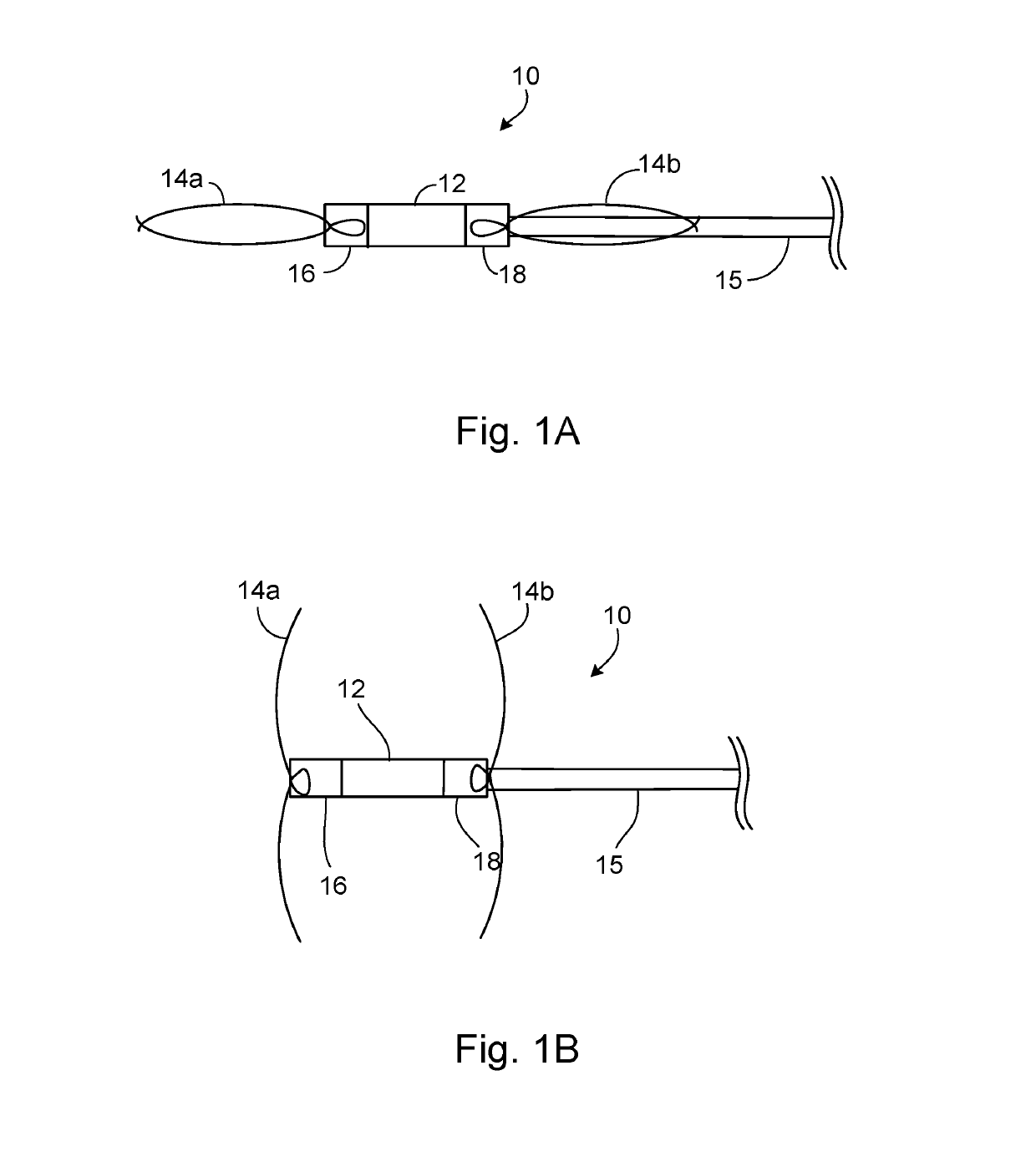
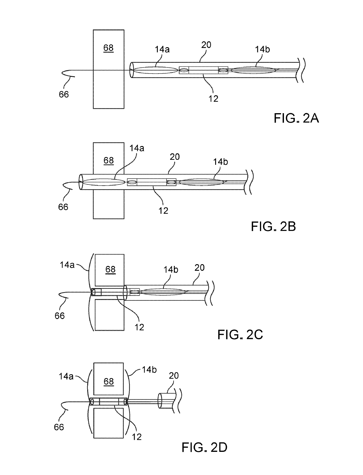
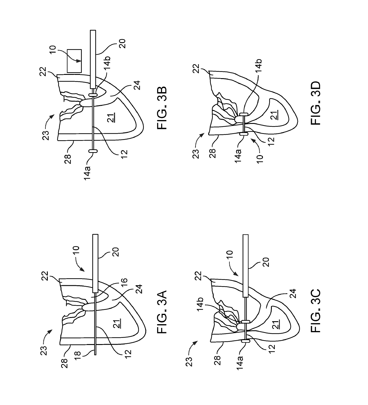
A device for treating tricuspid regurgitation includes a shaft and a first fixation mechanism disposed on a distal end of the shaft. The first fixation mechanism is configured to anchor the device to a first cardiac tissue. The device includes a second fixation mechanism disposed on a proximal end of the shaft. The second fixation mechanism is configured to anchor the device to a second cardiac tissue. The device includes an approximation mechanism connecting the first fixation mechanism and the second fixation mechanism, wherein the approximation mechanism causes the second fixation mechanism to move along the shaft toward the first fixation mechanism to approximate the first cardiac tissue and the second cardiac tissue.

Description

CROSS REFERENCE TO RELATED APPLICATIONS[0001]This application claims priority from U.S. Provisional Application Ser. No. 61 / 598,966, filed on Feb. 15, 2012, the contents of which are incorporated herein by reference in their entirety.STATEMENT AS TO FEDERALLY SPONSORED RESEARCH[0002]This invention was made with Government support under Grant No. HL073647 awarded by the National Institutes of Health. The Government has certain rights in the invention.FIELD OF THE INVENTION[0003]The invention relates to devices and methods for right ventricular papillary approximation.BACKGROUND OF THE INVENTION[0004]Tricuspid regurgitation (TR) adversely affects cardiac mortality in a number of clinical settings. In adults, “functional” TR from tricuspid annular dilation and right ventricular enlargement is a common TR mechanism and can be secondary to left-sided valve and ventricular dysfunction or pulmonary hypertension. Tricuspid annuloplasty (TAP) can be used to repair the tricuspid valve in case...

Claims


Application Information

Patent Timeline
Patent Type & Authority Patents(United States)
IPC IPC(8): A61F2/24A61B17/04A61M25/02A61B17/00A61B17/08A61M25/04
CPCA61F2/2487A61B17/0401A61B2017/0406A61B2017/0419A61B2017/081A61B2017/00407A61B2017/0409A61B2017/00243A61M2025/0233A61B2017/00606A61M25/04A61B2017/0464A61B2017/0451
Inventor DEL NIDO, PEDRO J.YAMAUCHI, HARUOVASILYEV, NIKOLAY V.NUNES PEREIRA, MARIA JOSE M.O'CEARBHAILL, EOIN D.
Owner THE BRIGHAM & WOMEN S HOSPITAL INC
Learn More