
Method and apparatus for selective treatment of biological tissue

a biological tissue and selective treatment technology, applied in the field of pigmented biological tissue, can solve the problems of ejection of tissue fragments from the tissue surface, mechanical (as well as thermal) disruption of nearby tissue, and small vapor bubble expansion, so as to facilitate selective energy absorption and avoid unwanted damage to surrounding areas. , the effect of sufficient selective absorption of local energy density

Active Publication Date: 2021-04-13
BLOSSOM INNOVATIONS LLC +1
View PDF8 Cites 2 Cited by
  • Summary
  • Abstract
  • Description
  • Claims
  • Application Information

AI Technical Summary

Benefits of technology

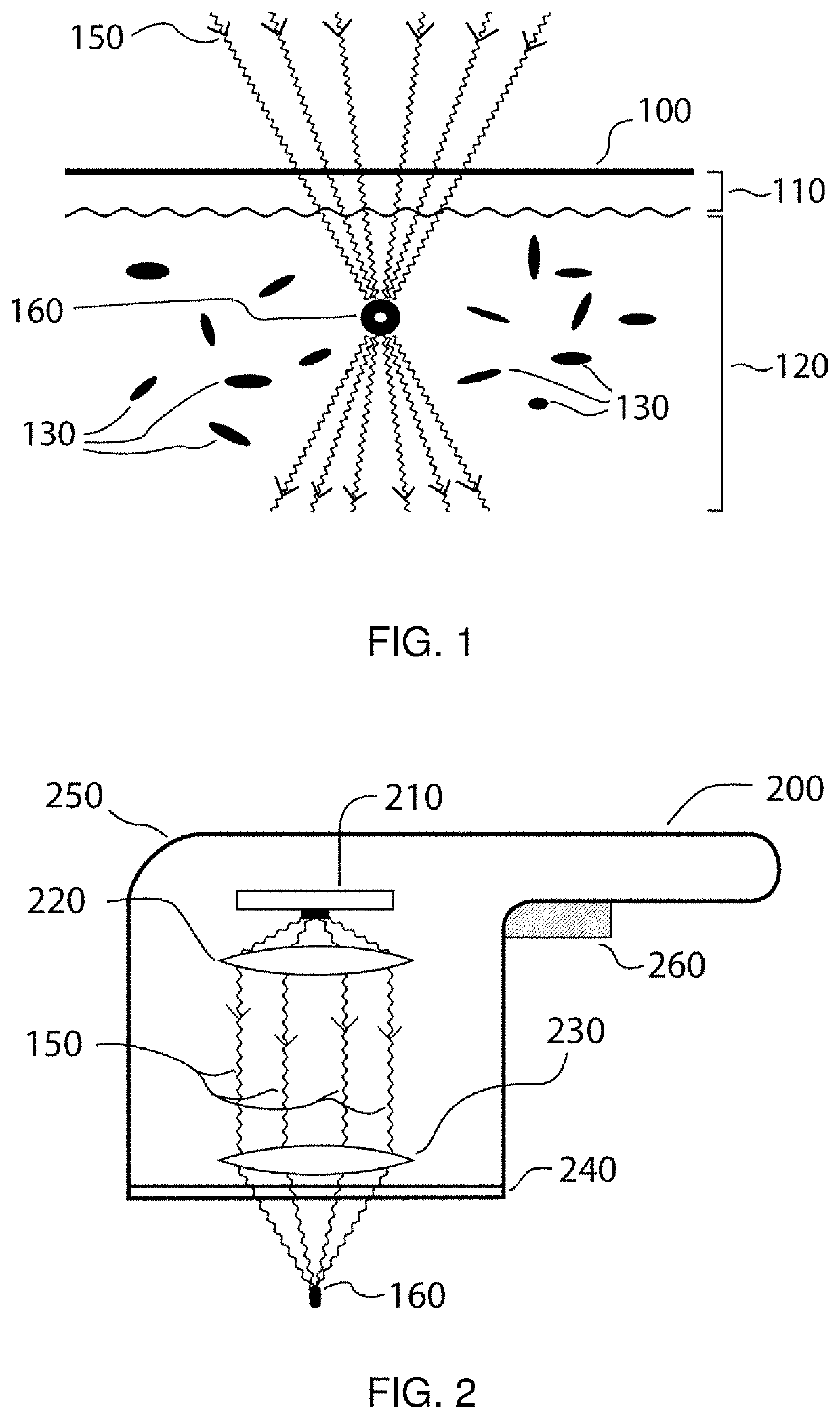
[0017]Exemplary embodiments of methods and apparatus can be provided for a treatment of biological tissue, for example, to selectively generate local plasma effects in pigmented regions of such tissue. The exemplary embodiments of the methods and apparatus can facilitate a selective energy absorption by pigmented or chromophore-containing structures and / or regions within biological tissues (e.g., skin tissue) by focusing highly-convergent electromagnetic radiation (EMR), e.g., optical energy, having appropriate wavelengths and other parameters onto regions within the tissue. This exemplary procedure can produce a sufficient selective absorption of local energy densities in the tissue to result in a production of plasmas in the biological tissue, e.g., arising from thermionic plasma initiation, which are selective to chromophore-containing tissue regions. Such localized plasmas can disrupt the pigment and / or chromophores while avoiding unwanted damage to surrounding unpigmented tissue and the overlying tissue. Such systems and methods described herein can be used, e.g., to improve appearance of skin tissue.
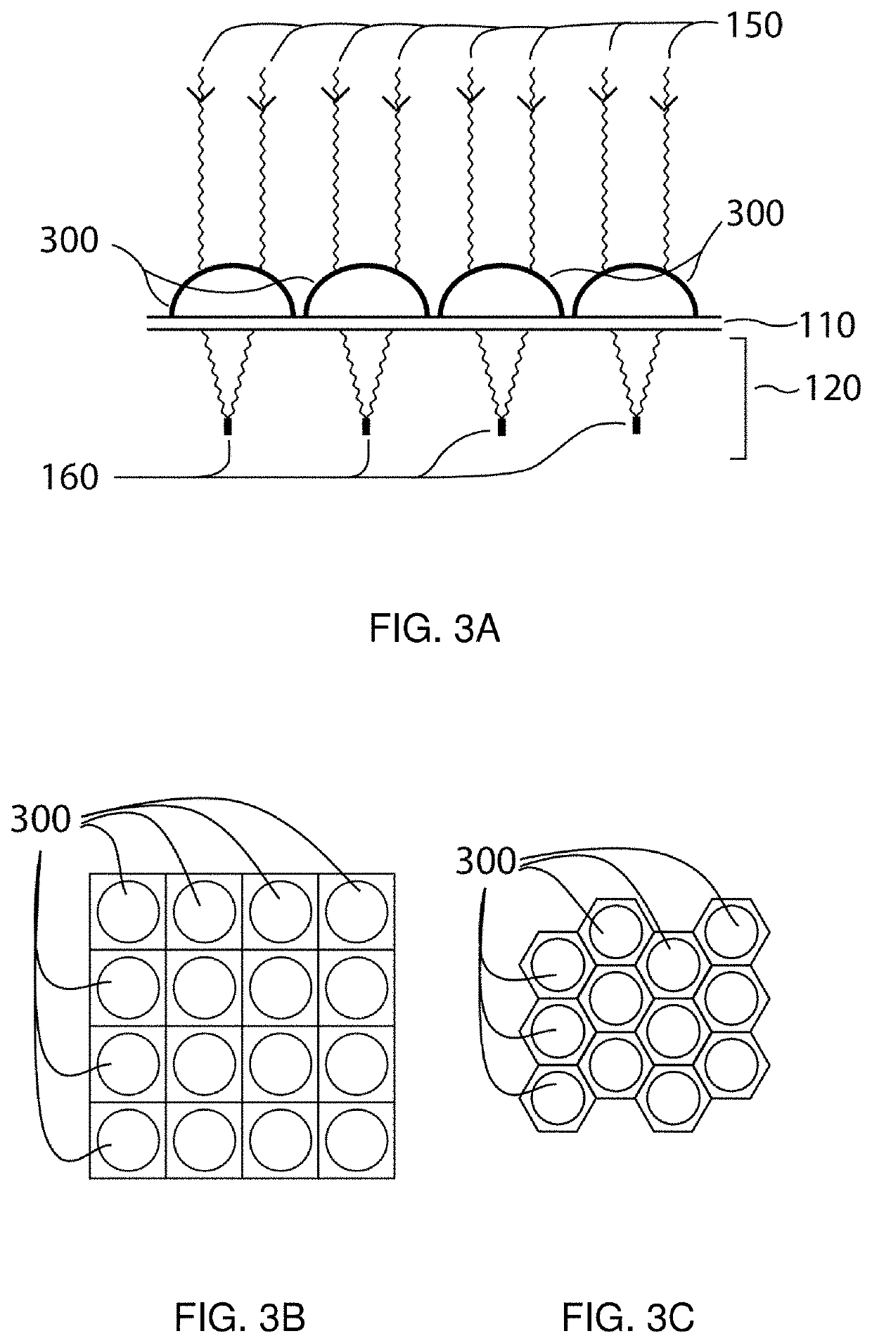
[0021]The exemplary apparatus can include an optical arrangement configured to focus the EMR in a highly convergent beam. For example, the optical arrangement can include a focusing or converging lens arrangement having a numerical aperture (NA) of about 0.5 or greater, e.g., between about 0.5 and 0.9. The correspondingly large convergence angle of the EMR can provide a high fluence and intensity in the focal region of the lens with a lower fluence in the overlying tissue above the focal region. Such focal geometry can help reduce unwanted thermal damage in the overlying tissue above the targeted tissue regions. The exemplary optical arrangement can further include a collimating lens arrangement configured to direct EMR from the emitting arrangement onto the focusing lens arrangement.
[0022]The exemplary apparatus can be configured to focus the EMR such that a local intensity or power density of the optical energy in the focal region is about 10{circumflex over ( )}10 W / cm2 or more, for example, between about 10{circumflex over ( )}10 W / cm2 and 10{circumflex over ( )}11 W / cm2 for optical energy having a wavelength of about 1060 nm. In certain embodiments, the local power density can be lower, e.g., as low as about 0{circumflex over ( )}8 W / cm2, if other parameters such as absorption efficiency (which depends in part on the chromophore and on wavelength of the optical energy) and energy density (which also depends in part on pulse duration) are selected appropriately. An optical arrangement can be provided to focus the EMR to a small spot size in the focal region, e.g., a spot size (as measured in air with reduced scattering) between about 5 μm and about 100 μm. Such small focal spot sizes can facilitate generation of sufficiently high local power densities in the foal region. Somewhat smaller or larger spot sizes can be used in certain exemplary embodiments, e.g., depending on other factors such as wavelength(s) of the optical energy and absorption coefficient by a particular chromophore at such wavelength(s).
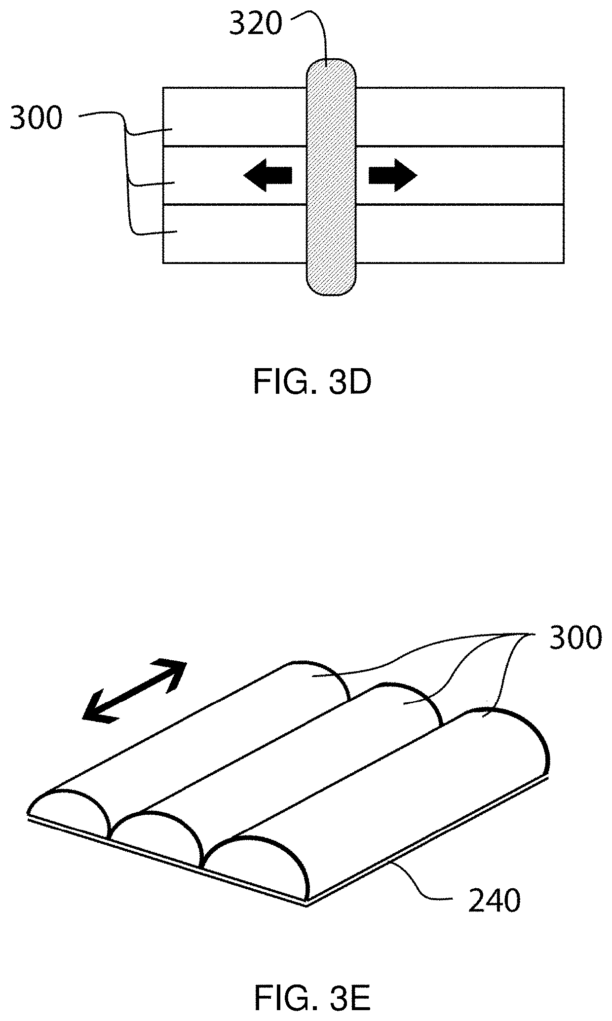
[0024]In further exemplary embodiments of the present disclosure, the positions and / or orientations of the EMR emitter arrangement and / or components of the optical arrangement can be controllable and / or adjustable relative to one another and / or relative to the tissue, such that the location and / or path of the focal region(s) in the tissue can be varied. Such variation in the path of the focal region(s) can be provided using optical arrangements having variable focal lengths, mechanical translators that can controllably vary the position of the optical arrangement and / or EMR emitter arrangement relative to the tissue being treated, etc. Such exemplary variations in location of the focal region(s) can facilitate treatment of larger volumes of the tissue by “scanning” the focal region(s) within the tissue, e.g., in a pattern at a particular depth and / or at multiple depths. In certain exemplary embodiments, a mechanical translator can be provided having scan speeds over an area of tissue to be treated that range from, e.g., about 5 mm / sec to about 5 cm / sec.
[0025]In further exemplary embodiments of the present disclosure, a handpiece can be provided that is configured to be manually translated over the tissue at similar speeds. Sensor arrangements can be provided in such manual handpieces or in mechanically-translated devices to detect scanning speeds and affect parameters of the EMR source (such as EMR pulse duration, pulse frequency, pulse energy, etc.) and / or optical arrangement based on such detection, e.g., to maintain a consistent range of parameters such as local power density and local dwell times during treatment. For example, scanning speeds and focal region spot sizes can be selected to maintain a sufficiently small local dwell time of the focal region at a location in the tissue (e.g. less than about 1-2 ms) to avoid damaging unpigmented tissue.

Problems solved by technology

Such photo ablation can produce rapidly-expanding small vapor bubbles in the tissue, which can generate mechanical (as well as thermal) disruption of nearby tissue, or ejection of tissue fragments from the tissue surface.
Selective or preferential absorption of the optical energy by chromophores can lead to local heating of the adjacent tissues, which can lead to thermal damage or necrosis of cells, physical changes in the heated tissue (e.g. coagulation, denaturation of collagen, etc.), and even vaporization of tissue.
In contrast, longer local irradiation times can lead to more widespread thermal damage arising from diffusion of heat away from the preferential absorption site.
Irradiation of tissue with high-intensity optical energy beams can also lead to dielectric breakdown of tissue components and formation of a plasma.
This can produce a chain reaction that results in a plasma formation, which is often accompanied by rapid local expansion and mechanical shockwaves in the tissue.
Because of the very high power densities and mechanisms of optical (or dielectric) breakdown, formation of plasma in tissue tends to be nonselective with respect to the presence of chromophores.

Method used


Image

  • Method and apparatus for selective treatment of biological tissue
  • Method and apparatus for selective treatment of biological tissue
  • Method and apparatus for selective treatment of biological tissue

Examples


example 1

[0126]An animal study using an exemplary spot-focused laser device and model system were used to test the efficacy of selective plasma formation in skin tissue using optical radiation. The study was performed on a female Yucatan pig, as described below.

[0127]First, a deep-melasma condition was simulated by tattooing the dermis using a melanin-based ink. The ink was prepared by mixing synthetic melanin at a concentration of 20 mg / mL in a 50:50 saline / glycerol solution. The resulting suspension was then agitated prior to being injected into approximately 1″×1″ test sites on the animal subject using a standard tattoo gun, at a depth range of about 200-400 μm. Each test site was provided with a darker black tattooed border using India ink to facilitate identification of the various test sites.

[0128]An exemplary melasma treatment system was constructed based on exemplary embodiments of the present disclosure described herein, which includes a Q-switched 1060 nm Yb-fiber laser with an ave...

example 2

[0136]FIG. 6 shows a further scanned melanin-tattooed test site that was irradiated using the general scan parameters indicated above (e.g., a scan rate of 200 mm / s, a repetition rate of 50 kHz, and six (6) sequential scanned layer depths of 550, 500, 450, 400, 350, and 300 m, and a distance between adjacent scan lines in each plane of 25 μm), with a fiber laser output of 1 W, at various times, in accordance with further embodiments of the present disclosure. Image 610 in FIG. 6 shows the test site just prior to a scanned irradiation using the laser apparatus, and image 612 shows the test site just after the scan was completed. Images 614, 616, 618, 620, and 622 illustrate the appearance of the test site 610 at 1 hour, 3 days, 1 week, 2 weeks, and 4 weeks, respectively, after the irradiation treatment. No plasma formation was observed at this lower power output level.

example 3

[0137]FIGS. 7A and 7B show images of a melanin-tattooed test site that was scanned twice, over two sessions spaced two weeks apart. Both irradiation treatments used a fiber laser with an average power output of 6 W and a pulse repetition rate of 20 kHz. The first scan session targeted more superficial layers (300 um to 550 um) whereas the second scan session targeted deeper layers (550 um to 850 um).

[0138]In particular, FIG. 7A illustrates exemplary results of the first scanned irradiation treatment. Image 710 in FIG. 7A shows the test site just prior to the first scanned irradiation using the laser apparatus, image 712 shows the test site just after the first scan was completed, and image 714 shows the test site 24 hours after the first scan was completed. Images 716, 718, and 720 provided in FIG. 7B show the appearance of the test site 710 just prior to, immediately following, and 24 hours following the second irradiation treatment, respectively. This second deeper irradiation tre...


PUM


Abstract

An exemplary treatment system can be provided which can include a laser system configured to emit at least one laser beam, and an optical system configured to focus the laser beam(s) to a focal region at a selected distance from a surface of a tissue. The focal region can be configured to illuminate at least a portion of a target. The optical system can cause an irradiation energy transferred to the focal region of the laser beam(s) to (i) generate a plasma in a first region of the tissue adjacent to the target, and (ii) avoid a generation of a plasma in a second region of the tissue. The optical system has a numerical aperture that is in the range of about 0.5 to about 0.9. An exemplary method can also be provided to control such treatment system.

Description

CROSS-REFERENCE TO RELATED APPLICATIONS[0001]The present application relates to and claims priority from U.S. Provisional Patent Application Ser. No. 62 / 438,818 filed Dec. 23, 2016, the disclosure of which is incorporated herein by reference in its entirety.FIELD OF THE DISCLOSURE[0002]Exemplary embodiments of the present disclosure relates to affecting pigmented biological tissue, and more particularly to methods and apparatus for selectively generating local plasma effects in pigmented regions of such tissue.BACKGROUND INFORMATION[0003]Affecting biological tissue with optical (light) energy has gained widespread use over the past few decades. Optical energy is a form of electromagnetic energy. In the electromagnetic spectrum, optical energy can typically range from the infrared regime (longer wavelengths) to the ultraviolet regime (shorter wavelengths). Treatment of biological tissue with optical energy typically involves introducing the optical energy into the tissue.[0004]When o...

Claims


Application Information

Patent Timeline
Patent Type & Authority Patents(United States)
IPC IPC(8): A61B18/20A61B18/00
CPCA61B18/203A61B2018/00452A61B2018/00577A61B2018/00625A61B2018/00642A61B2018/00696A61B2018/00708A61B2018/20355A61B2018/20553A61B2018/00732A61B18/042A61B2018/00702
Inventor ANDERSON, RICHARD ROXMANSTEIN, DIETERCHAN, HENRY HIN LEEZUO, VINCENT
Owner BLOSSOM INNOVATIONS LLC
Learn More