[0006]Embodiments therefore provide a medical device for occluding abnormal openings in a patient's vasculature. In general, the medical device is configured such that an end feature of the device is recessed within a tapered transition portion formed at a respective end of the medical device. In this way, only an end surface of the end feature (e.g., a proximal end surface of the end feature at the proximal end of the medical device), or a portion of this surface, is exposed to the flow of blood through the body lumen, and tissue in-growth over the end of the device may be enhanced and facilitated. Further, the medical device of the present disclosure may include a distal end feature that is sized and configured to increase the potential for a more atraumatic delivery of the medical device (i.e., decrease the potential of possible tissue damage during delivery of the medical device) by providing a more atraumatic leading edge of the medical device.
[0007]In addition, the medical device is configured to include a plurality of stabilizing wires or retention members that are configured to be at least partially retractable, and desirably substantially retractable, within the medical device when the medical device is in a stretched, elongated, or collapsed configuration. By configuring the stabilizing wires in this manner, interaction between the stabilizing wires and the inner surface of a delivery catheter or delivery sheath is reduced, which in turn may reduce the potential for damage to the inner surface of the delivery catheter or delivery sheath by the stabilizing wires. That is, by configuring the stabilizing wires in this manner, the ability of the medical device to smoothly pass through the delivery catheter or sheath is improved, thus reducing potential interaction with and damage to the delivery catheter or sheath during delivery and / or repositioning. This may also increase the number of times or the extent to which the medical device may be recaptured and redeployed (e.g., repositioned) within a single procedure. Further, by configuring the stabilizing wires such that they are at least partially, and desirably substantially, retracted within the medical device when in a stretched, elongated, or collapsed configuration, the force required by a user to recapture the device within the delivery catheter or sheath may be reduced due to the reduced interaction between the stabilizing wires and the delivery catheter or sheath (i.e., the inner surface or the distal end thereof).
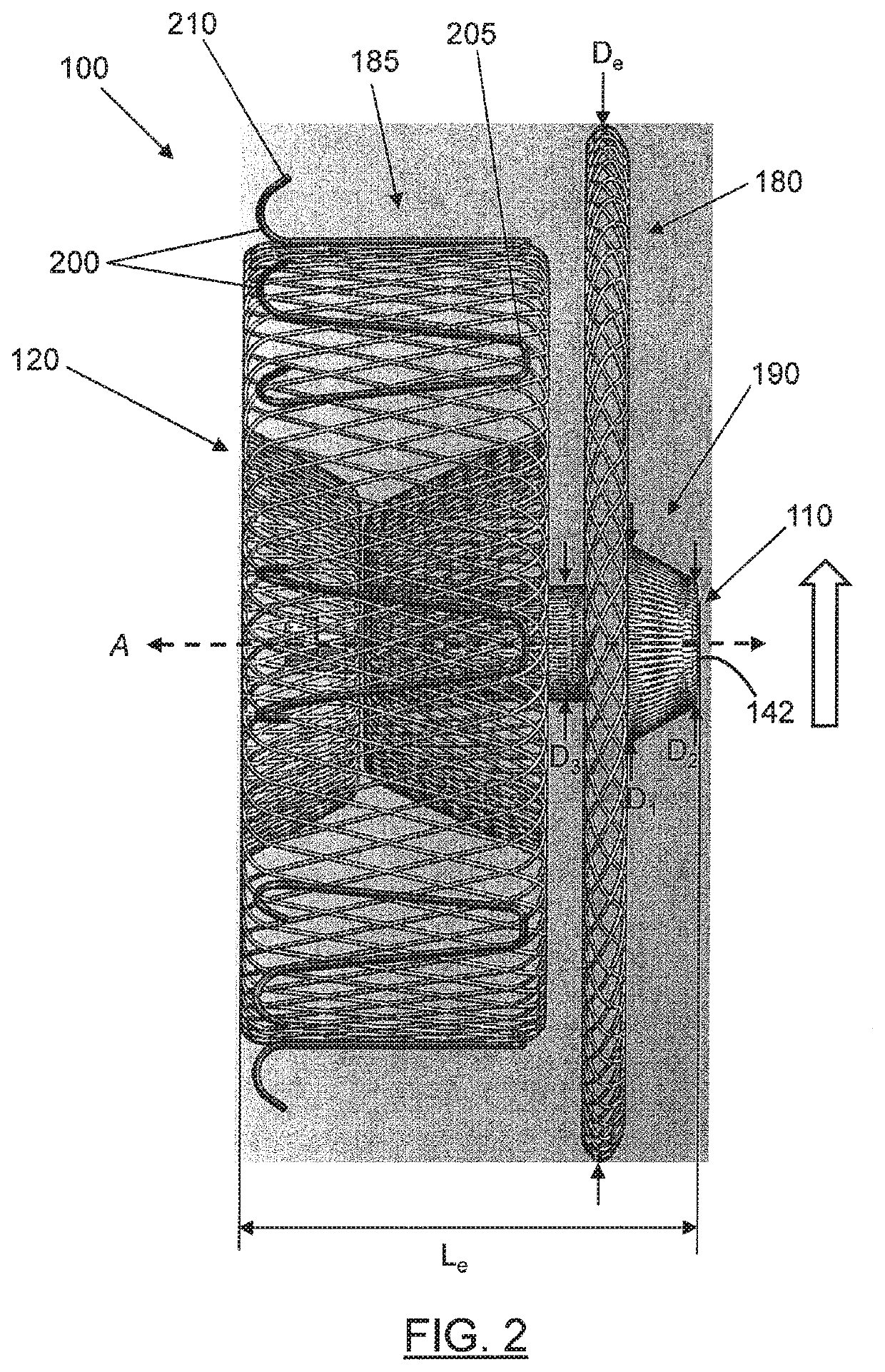
[0009]In one embodiment, a device is provided that is configured to self-expand from a contracted state when constrained within a delivery device toward an expanded state when deployed from the delivery device for delivery to a target site within the body lumen. The medical device may include a tubular structure and a first end feature. The tubular structure may comprise a plurality of braided strands, with each braided strand comprising a proximal strand end and a distal strand end. The first end feature may define a proximal end and a distal end, and the first end feature may be configured to receive and secure the proximal strand ends via the proximal end of the first end feature. The tubular structure may comprise an expanded volume portion proximate to the first end feature and a tapered transition portion extending between the expanded volume portion and the proximal end of the first end feature. In the expanded state, the expanded volume portion of the tubular structure may define an expanded volume diameter. Moreover, in the expanded state, the tapered transition portion may define a first transition diameter proximate the expanded volume portion and a second transition diameter proximate the proximal end of the first end feature. The first transition diameter may be greater than the second transition diameter, smaller than the expanded volume diameter, and disposed between the second transition diameter and the expanded volume diameter. In addition, the second transition diameter may be substantially equal to a diameter of the first end feature. In some cases, the second transition diameter may be sized to facilitate tissue growth over a proximal end of the medical device.
[0010]Embodiments of the medical device may also include a second end feature configured to receive and secure the distal strand ends of the plurality of braided strands. The medical device may define a central axis extending between the first end feature and the second end feature, and the expanded volume portion may define at least one surface that is substantially perpendicular to the central axis. In some cases, the expanded volume portion may define two surfaces that are substantially perpendicular to the central axis. The second end feature may define a proximal end and a distal end, and in one embodiment, the second end feature may be configured to receive and secure the distal strand ends via the distal end of the second end feature. In another embodiment, the second end feature is configured to receive and secure the distal end strands via the proximal end of the second end feature. In this embodiment, the second end feature is sized and configured to increase the potential for a more atraumatic delivery of the device (i.e., reduce the potential for tissue damage during delivery of the medical device). That is, the second end feature may include a smooth-radiused, or mushroom-shaped distal portion and a narrower proximal portion such that the potential for tissue trauma or damage during insertion of the distal portion of the second end feature through the vasculature and / or into the occlusion site may be reduced. In other words, the second end feature may be sized and configured to provide for a leading edge of the device that has a reduced potential for tissue damage upon delivery of the device.
[0011]In some cases, the expanded volume portion may be a first expanded volume portion and the tapered transition portion may be a first tapered transition portion. The tubular structure may further include a second expanded volume portion displaced from the first expanded volume portion and proximate the second end feature. In one embodiment, the device may include a second tapered transition portion extending between the second expanded volume portion and the distal end of the second end feature. In another embodiment, the second expanded volume portion may be substantially cylindrical and include a second end feature configured to reduce the potential for tissue trauma during insertion of a distal portion of the second end feature through the vasculature and / or into the occlusion site.
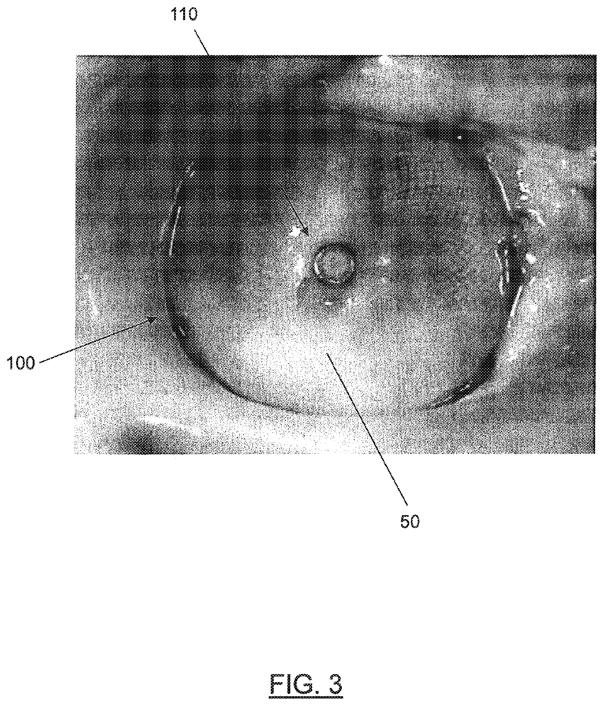
[0015]In some embodiments, the medical device may define a proximal end and a distal end, and the proximal end of the first end feature may substantially coincide with the proximal end of the medical device. The medical device may be configured to occlude a vessel, cavity, hole, septal defect, or lumen in a body. For example, the medical device may be configured to occlude the left atrial appendage of the heart and to prevent thrombus from escaping therefrom.

