Surgical clip and deployment system

a surgical clip and clip technology, applied in the field of surgical clips, can solve the problems of limited clip size, added time and complexity of surgical procedures, and limited jaw rang

Active Publication Date: 2022-08-16
THERAGI LLC
View PDF58 Cites 0 Cited by
  • Summary
  • Abstract
  • Description
  • Claims
  • Application Information

AI Technical Summary

Benefits of technology

[0006]The present invention overcomes the problems and deficiencies of the prior art. The present invention advantageously provides a clip for compressing tissue which can be controllably moved to the open position. The clip can also in some embodiments be controllably moved to the closed position. The clip can further advantageously be removed and repositioned after placement on body tissue. The present invention also provides a system and method for delivering the clip which is easily mountable over an endoscope and provides controlled manipulation.
[0036]In some embodiments, the method further comprises the step of retracting the sheath to increase the field of vision of the endoscope. In some embodiments, the endoscope can be advanced through the opening in the open clip.

Problems solved by technology

However, this approach has the disadvantage of the size of the clip being limited since it must be dimensioned to fit through the small dimensioned working channel of the scope.
If the clip delivery instrument has jaws, the range of the jaws is limited due to the size limitations of the working channel.
With the size restrictions, the clip in certain applications is unable to fully clamp the vessel or tissue, resulting in insufficient tissue clamping and / or requiring multiple clips to be applied which adds to the time and complexity of the surgical procedure.
First, there is no controlled opening or closure of the clip since the clip is biased open by the endoscope cap and springs back to a closed position when deployed off the cap.
Second, due to the positioning of the clip, visualization is compromised.
Third, once the clip is released onto tissue, it cannot be re-opened and repositioned.

Method used


Image

  • Surgical clip and deployment system
  • Surgical clip and deployment system
  • Surgical clip and deployment system

Examples


Embodiment Construction

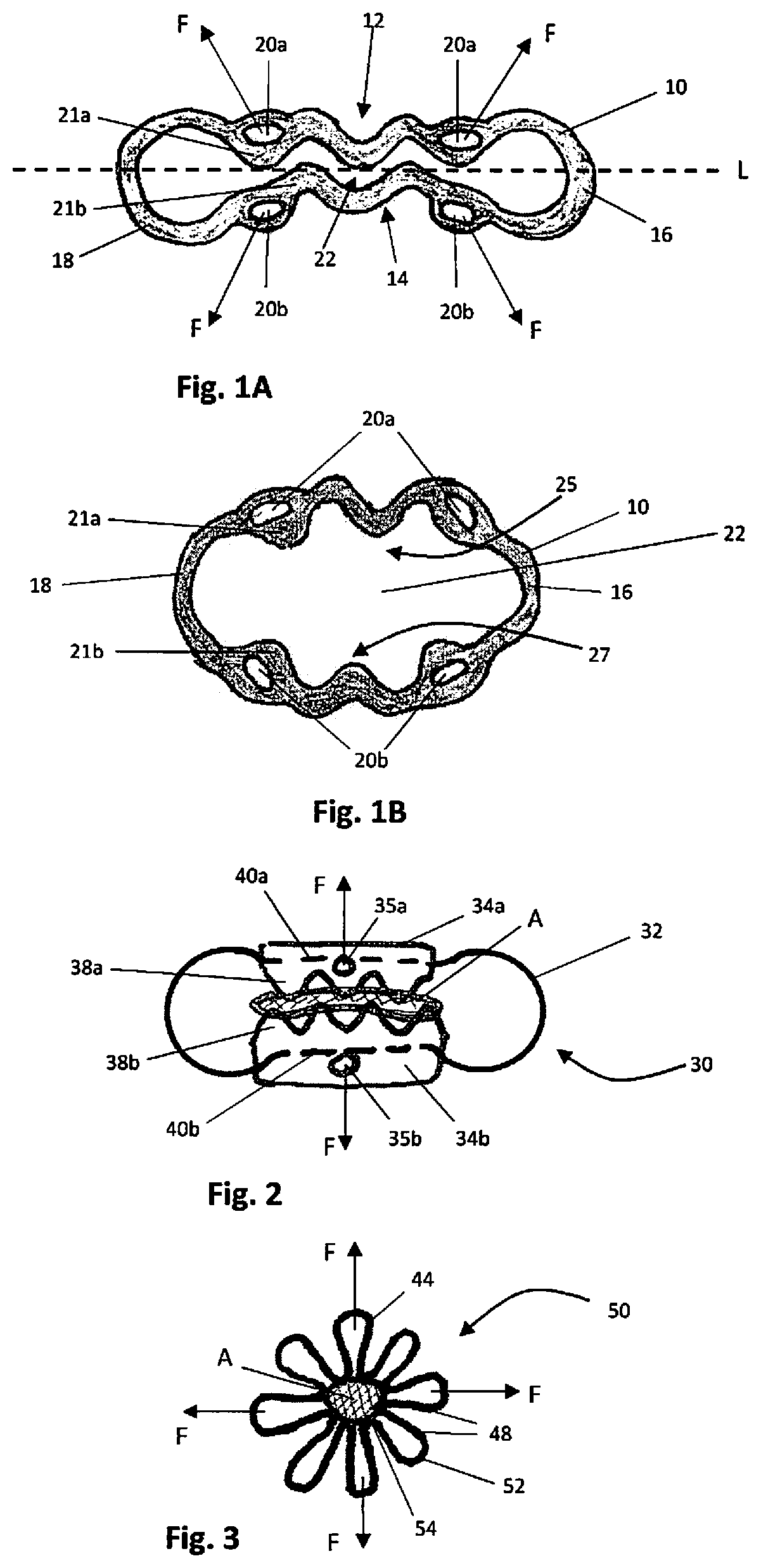
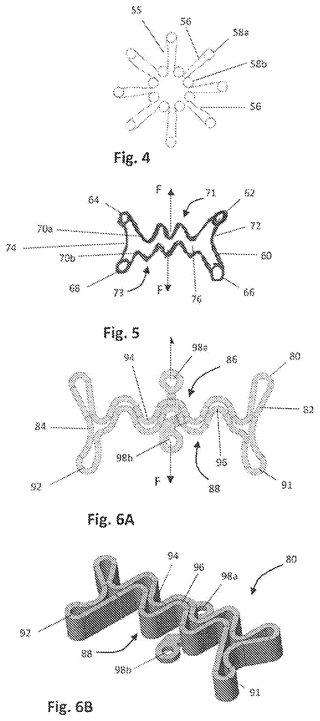
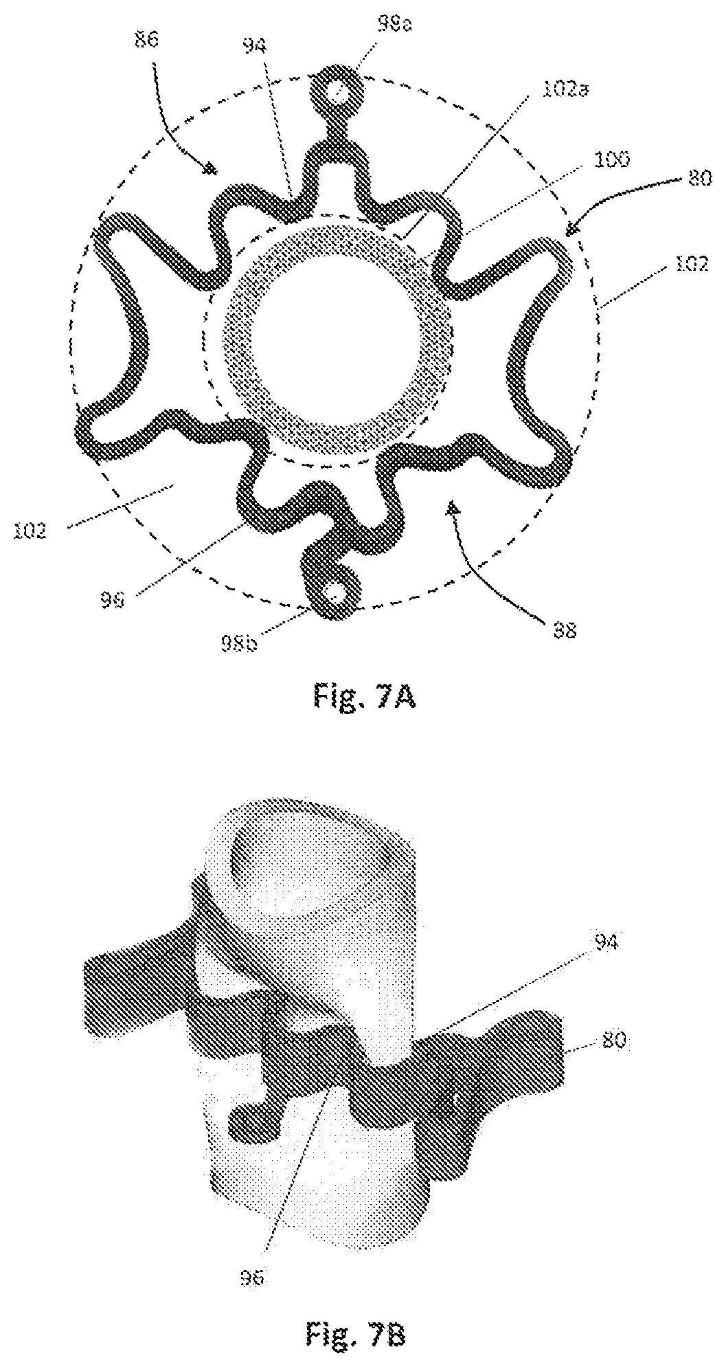
[0103]The present invention provides a system and method for closure of wall defects in the hollow organs, such as a colon, esophagus, stomach etc. The system includes a surgical clip and a deployment device for delivering the surgical clip to tissue and manipulating the clip between closed and open positions by applying a force to opposing sides of the clip. In one approach / aspect, the clips of the present invention are radially expandable from a closed position to an open position to enable tissue to be positioned within an opening in the clip, and then returnable to the closed position to compress tissue between opposing compression surfaces or points of the clip. Various embodiments of the radially expandable clips are discussed in detail below. The opening of the clips is controlled by a clip deployment device which has clip engagement members actuable by the clinician outside the patient, such actuation applying a force to opposing sides of the clip to spread the tissue contac...


PUM


Abstract

A system for compressing body tissue including a clip having first and second tissue contacting surfaces. The clip is movable from a closed position to an open position wherein the clip receives tissue between the first and second tissue contacting surfaces to compress tissue between the first and second tissue contacting surfaces. A clip deployment device has a first clip engagement member and a second clip engagement member engageable with the clip, the first and second clip engagement members movable between first and second positions to controllably move the clip from the closed position to the open position.

Description

[0001]This application is a 371 of International PCT / US18 / 67432, filed on Dec. 23, 2018, which claims priority to provisional application 62 / 613,902, filed Jan. 5, 2018, provisional application 62 / 648,586, filed Mar. 27, 2018, and provisional application 62 / 648,593, filed Mar. 27, 2018. The entire contents of each of these applications are incorporated herein by reference.BACKGROUND OF THE INVENTION1. Field of the Invention[0002]This application relates to a surgical clip and a system and method for deploying the surgical clip to compress tissue.2. Background of the Related Art[0003]Clips for closing defects in the GI tract are known. In one approach, clips are inserted through a working channel in an endoscope. However, this approach has the disadvantage of the size of the clip being limited since it must be dimensioned to fit through the small dimensioned working channel of the scope. Additionally, the instrument for delivering the clip must also be of small diameter since it also...

Claims


Application Information

Patent Timeline
Patent Type & Authority Patents(United States)
IPC IPC(8): A61B17/122A61B17/128
CPCA61B17/1227A61B17/1285A61B17/10A61B17/083A61B2017/00296
Inventor SHIKHMAN, OLEGRADZIUNAS, JEFFREYLAMOTHE, BRIAN MICHAELGOLDENBERG, CAROLLYNN
Owner THERAGI LLC
Why Eureka
Try Eureka