
Virus clearance of neoplastic cells from mixed cellular compositions

a technology of neoplastic cells and viruses, applied in the direction of dsrna viruses, drug compositions, tumor/cancer cells, etc., can solve the problems of cancer reappearing shortly after chemotherapy termination, cancer cells contaminated with cancer cells, and doses which are harmful to cancer cells are often harmful to thc hematopoietic stem cells

Inactive Publication Date: 2005-02-03
ONCOLYTICS BIOTECH
View PDF19 Cites 13 Cited by
  • Summary
  • Abstract
  • Description
  • Claims
  • Application Information

AI Technical Summary

Benefits of technology

[0053] In another embodiment of the present invention, the virus is removed from the virus-treated cellular composition by using a gradient which can separate viruses from cells.
[0059] In another aspect of this invention, the virus selectively kills neoplastic cells by carrying a tumor suppressor gene. For example, p53 is a cellular tumor suppressor which inhibits uncontrolled proliferation of normal cells. Approximate half of all tumors have functionally impaired p53 and proliferate in an uncontrolled manner. Therefore, a virus which expresses the wild type p53 gene can selectively kill the neoplastic cells which become neoplastic due to inactivation of the p53 gene product.

Problems solved by technology

Therefore, any dosage which is harmful to cancer cells is often also harmful to thc hematopoietic stem cells.
On the other hand, if the dosage is not high enough to kill the cancer cells, there is a risk that the cancer would reappear shortly after chemotherapy is terminated.
A serious drawback of this therapy is that when the hematopoietic progenitor stem cells are removed from the patients, they are often contaminated with cancer cells.
This is especially a problem when the patient has a cancer of hematopoietic origin, but patients with a solid tumor may also suffer from contamination of the hematopoietic stem cells, particularly if the solid tumor has metastasized.
However, as discussed above, it is hard to find a dosage for the chemotherapeutic drug which selectively kills neoplastic cells or cancer cells but leaves normal hematopoietic stem cells intact.
However, by selecting only certain hematopoietic cells, e.g., the CD34+ cells, other hematopoietic cells such as T cells, B cells, monocytes and natural killer cells are also eliminated, and immune recovery may be delayed (Bensinger, 1998).
This method also results in the loss of about half the CD34+ cells and retention of some contaminating cancer cells (Spyridonidis et al., 1998).

Method used


Image

  • Virus clearance of neoplastic cells from mixed cellular compositions
  • Virus clearance of neoplastic cells from mixed cellular compositions
  • Virus clearance of neoplastic cells from mixed cellular compositions

Examples


example 1

Reovirus Induced Oncolysis and Apoptosis in Breast Cancer Cells

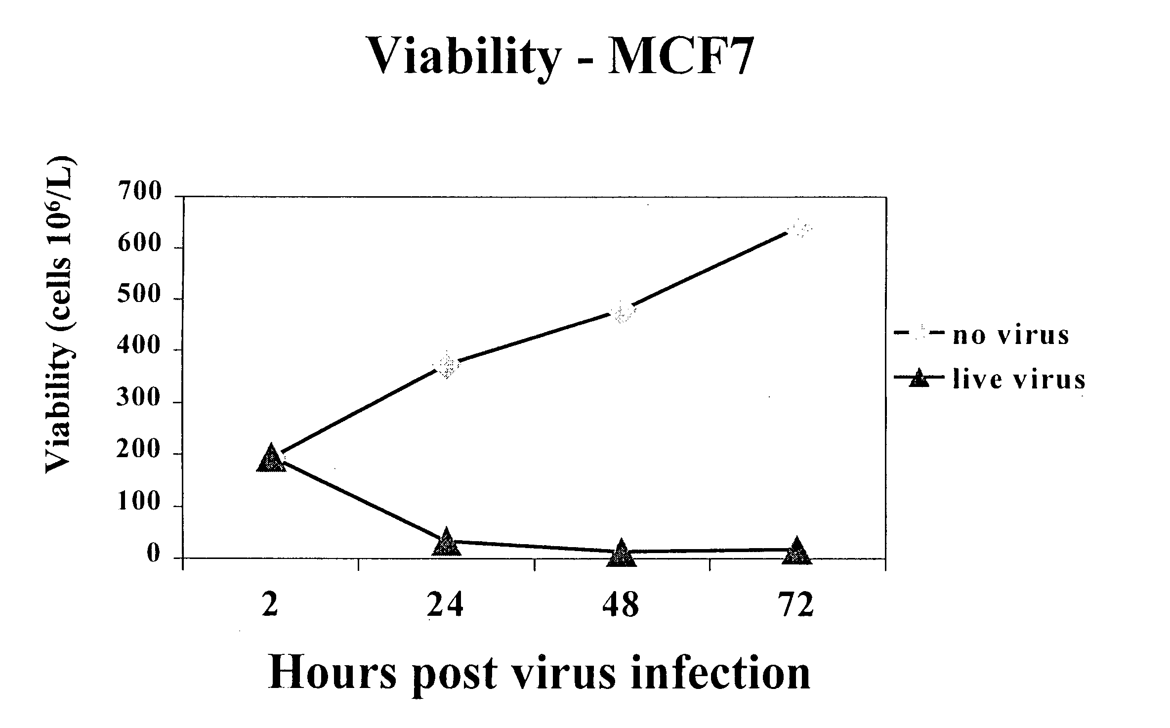
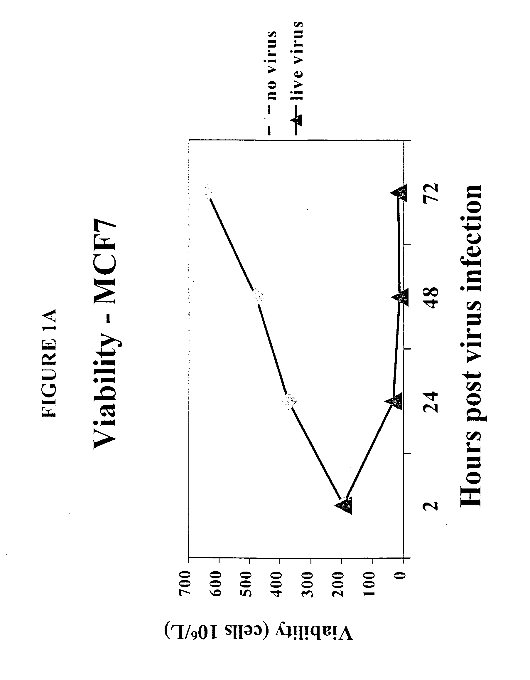
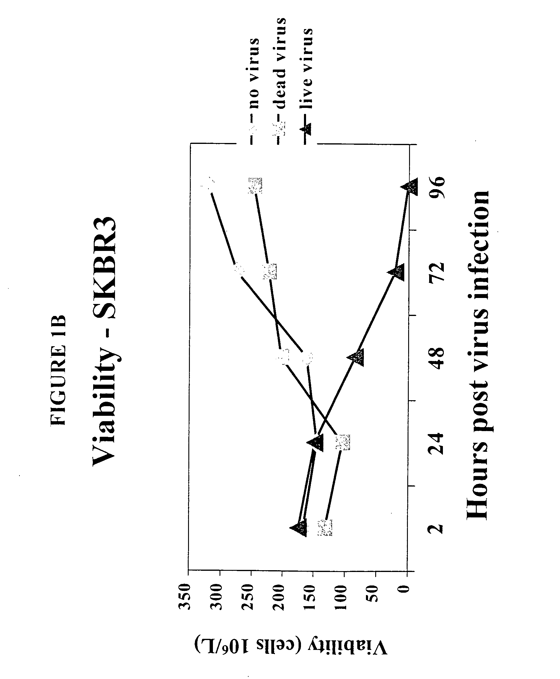
[0149] To determine the effect of reovirus on the viability of neoplastic cells, we first used three breast cancer model systems, MCF7 (ATCC number HTB-22), SKBR3 (ATCC number HTB-30) and MDA MB 468 (ATCC number HTB 132). Cells of each cell line were grown to 50-60% confluency and infected with reovirus serotype 3, strain Dearing, at a multiplicity of infection of 40. Reovirus was obtained and maintained as described in U.S. Pat. No. 6,136,307. Reovirus infected and non-infected cells were harvested at 0, 24, 48 and 72 hours after infection and the viability was determined.

[0150] The results are shown in FIGS. 1A-1D. Viable cell count in reovirus-infected MCF7 (FIG. 1A), SKBR3 (FIG. 1B) or MDA MB 468 cells (FIG. 1C) dropped significantly after the infection, while the cells infected with dead virus or no virus. proliferated as expected. Reovirus treatment caused MCF7 (FIG. 1D) and SKBR3 viability to drop from 93% to 16...

example 2

Reovirus Selectively Inhibited Protein Synthesis in Cancer Cells but not CD34+ Stem Cells

[0152] For further proof of selective viral infection of cancer cells, 35S labeling / SDS / PAGE of viral proteins was undertaken. Viral protein synthesis was evident after 1-2 days in MCF7 cells infected with reovirus, while cellular protein synthesis decreased at the same time, indicating that reovirus had taken over the cellular machinery. At 4 days after infection, no protein synthesis could be detected anymore, suggesting that all the cells had been killed. In the control experiments where cells were infected with dead reovirus or no virus, there was no viral protein synthesis, whereas cellular protein synthesis was at the normal level. In contrast, 35S labeling of CD34+ stem cells in the presence or absence of reovirus showed no viral protein synthesis up to 72 hours after the addition of virus. Therefore, reovirus selectively infect MCF7 cells but not CD34+ stem cells.

example 3

Reovirus Treatment Neither Inhibited Cell Proliferation nor Altered Differentiation Potential of CD34+ Cells

[0153] Consistent with the protein synthesis results, viable cell count indicated that reovirus treatment did not decrease the number of viable cells in CD34+ cells (FIG. 3A) as compared to the no virus control.

[0154] While the number of CD34+ cells was unaffected by reovirus infection, there remained the question whether reovirus changed the potential of CD34+ stem cells to differentiate into all the hematopoietic lineages in the appropriate proportion. If this was the case, reovirus treated stem cells would not be a good candidate for the reconstitution of the whole hematopoietic system. To investigate this possibility, CD34+ cells were incubated with reovirus for 2, 24, 48 or 72 hours, respectively. The reovirus was then removed and the cells were diluted and cultured in fresh media for 14 days to allow colonies to form. Each colony was examined to determine if it belongs...


PUM

PropertyMeasurementUnit
Compositionaaaaaaaaaa

Abstract

The present invention relates to a method for removing neoplastic cells from a mixed cellular composition, which is outside of a living organism, by using a virus which selectively infect and kill neoplastic cell. A variety of viruses can be used in this method to remove neoplastic cells for different purposes, for example, to purge hematopoietic stem cells prior to transplantation. Also provided are compositions prepared according to this method, and kits comprising a combination of viruses which are useful in this invention.

Description

RELATED INVENTIONS [0001] This application claims the benefit of U.S. Provisional Applications Ser. No. 60 / 201,990, filed May 3, 2000, Ser. No. 60 / 205,389, filed May 19, 2000, Ser. No. 60 / 268,054, filed Feb. 13, 2001 and Ser. No. 60 / 276,782, filed Mar. 16, 2001, under 35 U.S.C. §119(e). The entire disclosure of each of the above provisional applications is hereby incorporated by reference.FIELD OF THE INVENTION [0002] The present invention relates to a method of selectively removing neoplastic cells from a mixed cellular composition outside of a living organism by using a virus which selectively infects and kills the neoplastic cells. Also provided are compositions prepared according to this method, and kits comprising a combination of viruses which are useful in this invention. References [0003][0003] U.S. Pat. No. 6,136,307. [0004] WO 94 / 18992, published Sep. 1, 1994. [0005] WO 94 / 25627, published Nov. 10, 1994. [0006] WO 99 / 08692, published Feb. 25, 1999. [0007] Bar-Eli, N., et a...

Claims


Application Information

Patent Timeline
IPC IPC(8): A61K35/12A61K35/14A61K35/23C12N7/00A61K35/28A61K35/34A61K35/36A61K35/38A61K35/39A61K35/407A61K35/52A61K35/54A61K35/76A61K35/765A61K48/00A61L2/00A61P43/00C12N5/00C12N5/09C12N15/861
CPCA61K48/00A61L2/00C12N5/0093C12N5/0693A61K35/28C12N2500/70C12N2720/12232C12N2720/12243A61K35/765C12N15/86A61P35/00A61P35/02A61P43/00
Inventor MORRIS, DONALDTHOMPSON, BRADLEY G.COFFEY, MATTHEW C.
Owner ONCOLYTICS BIOTECH
Learn More