Systems and methods for providing automated regional myocardial assessment for cardiac imaging

a cardiac imaging and regional myocardial technology, applied in the field of systems and methods for providing automated regional myocardial assessment for cardiac imaging, can solve the problems of occlusion of cardiac arteries, requiring invasive procedures, and unable to measure or otherwise assess the effects of such occlusions

Active Publication Date: 2005-03-17
SIEMENS MEDICAL SOLUTIONS USA INC
View PDF14 Cites 218 Cited by
  • Summary
  • Abstract
  • Description
  • Claims
  • Application Information

AI Technical Summary

Benefits of technology

In another exemplary embodiment of the invention, a method for automatically assessing the condition of myocardial tissue of one or more regions of the myocardial wall is performed using a classification method that is trained to automatically classify the condition of myocardial tissue for one or more regions of the myocardial wall. The method for automatically classifying can be implemented using a machine learning methods, model-based methods, or any combination of machine learning and model-based methods.
In yet another embodiment of the invention, the process of automatically classifying the condition of myocardial tissue of one or more regions of the myocardial wall is performed by obtaining features for each of a plurality of pre-defined segments of a myocardial wall and automatically classifying a condition of myocardial tissue for each segment of the myocardial wall. In one embodiment, the condition of myocardial tissue for each segment of the myocardial wall is classified using an indicator that indicates whether the myocardial tissue for each segment is normal or abnormal. For example, in one exemplary embodiment of the invention, the indicator comprises a wall motion score that is automatically generated for each segment in accordance with a standard specified by the ASE (American Society of Echocardiography). In yet another embodiment of the invention, a measure of confidence is determined for each classified condition of myocardial tissue for each segment of the myocardial wall.

Problems solved by technology

Coronary artery disease and other heart-related diseases are very prevalent, especially in western civilizations, and lead to the death of many people each year.
However, these measurements often require invasive procedures.
Furthermore, although angiography can be used to identify and measure occlusions, such methods cannot measure or otherwise assess the effects of such occlusions.
Indeed, the effect of coronary occlusion is typically manifested regionally within the heart wall, resulting in abnormalities of myocardial tissue or myocardial function.
For example, one conventional method includes automated border detection based on analysis of integrated backscatter, which provides an automated estimate of LV function indices, but does not address segmental or global wall function.
Other methods for automatic wall motion analysis generate parametric images indicating excursion, but provide no quantitative comparison amongst segments.
While automated methods for wall motion analysis can provide parametric images and generate indices related to wall motion, such methods do not provide automated assessment, or otherwise identify or cnaracterize the condition (e.g., normal or abnormal) of the myocardial tissue.

Method used


Image

  • Systems and methods for providing automated regional myocardial assessment for cardiac imaging
  • Systems and methods for providing automated regional myocardial assessment for cardiac imaging
  • Systems and methods for providing automated regional myocardial assessment for cardiac imaging

Examples


Embodiment Construction

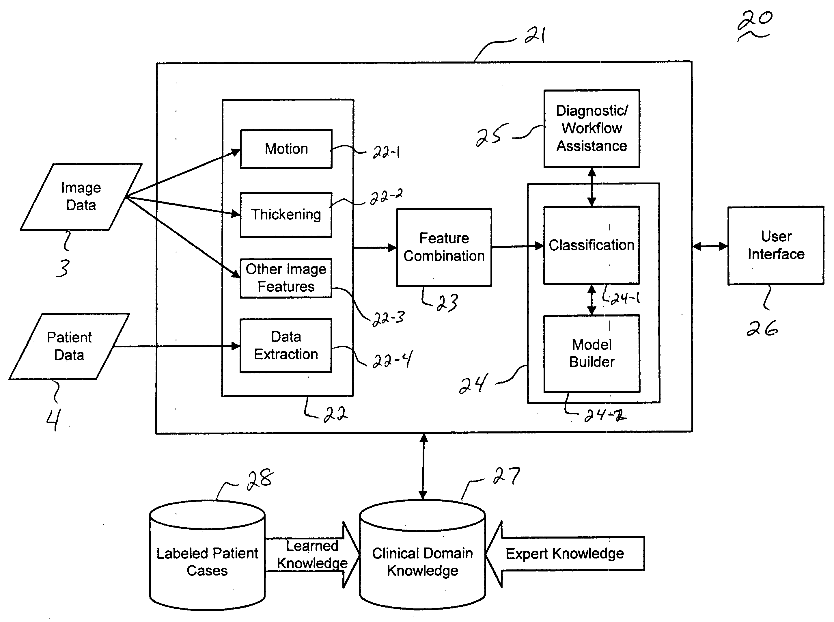
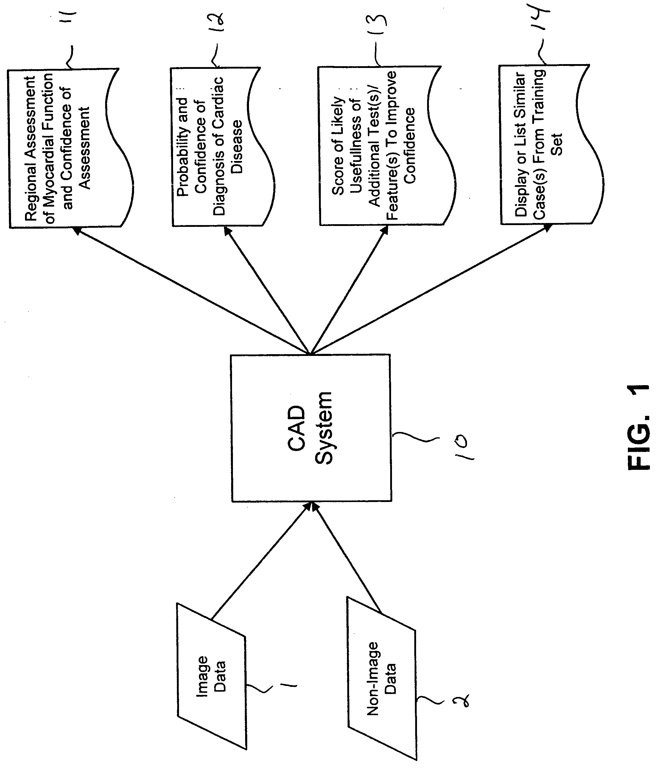
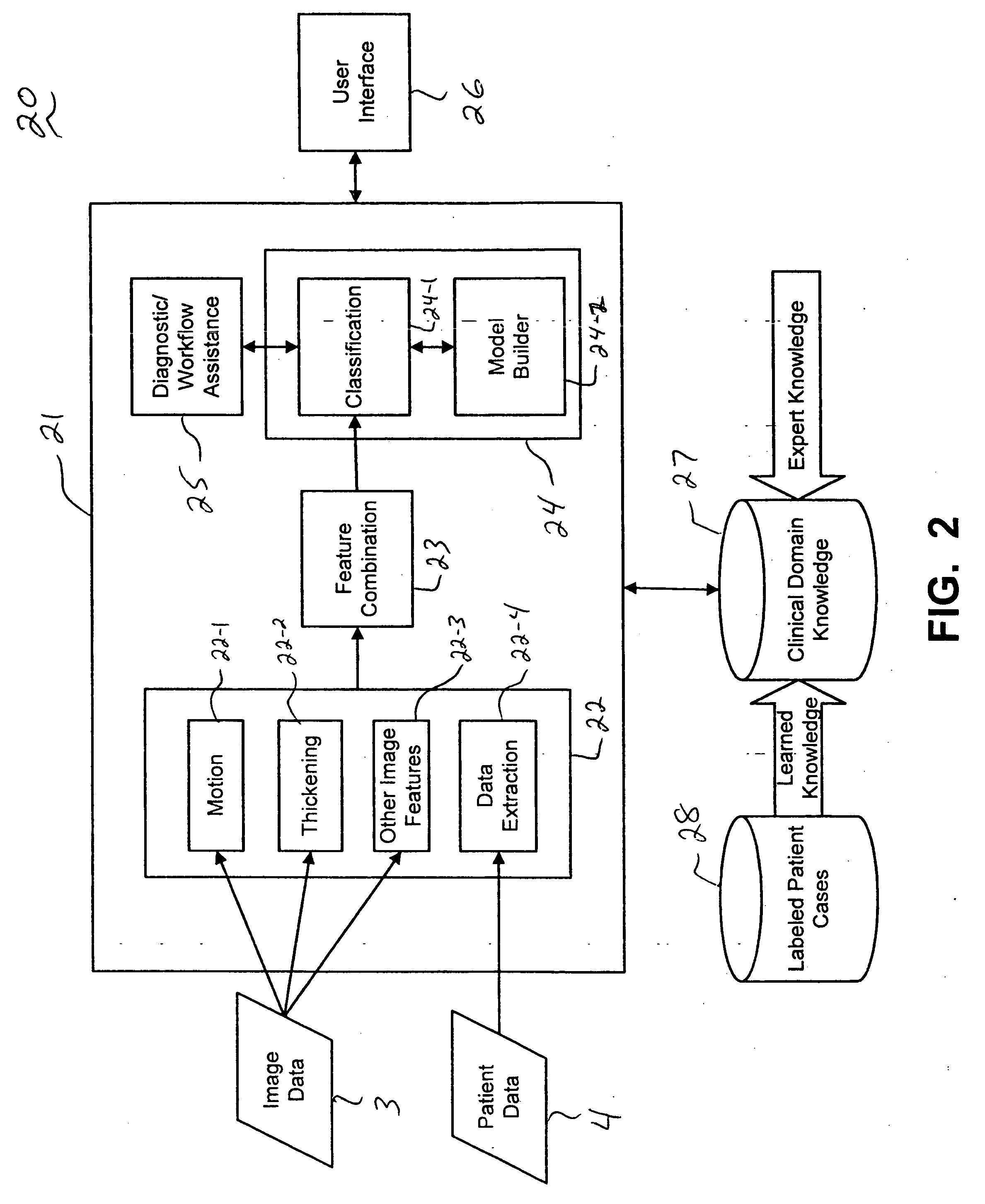
In general, exemplary embodiments of the invention as described below include systems and methods for providing automated diagnosis and decision support for cardiac imaging. More specifically, exemplary embodiments of the invention as described below with reference to FIGS. 1˜4, for example, include CAD (computer-aided diagnosis) systems and applications for cardiac imaging, which implement automated methods for extracting and analyzing relevant features / parameters from a collection of patient information (including image data and / or non-image data) of a subject patient to provide automated assistance to a physician for various aspects of physician workflow including, for example, automated assessment of regional myocardial function through wall motion analysis, automated diagnosis of heart diseases and conditions such as cardiomyopathy, coronary artery disease and other heart-related medical conditions, and other automated decision support functions to assist physician workflow. T...


PUM


Abstract

Systems and methods are provided for automated assessment of regional myocardial function using wall motion analysis methods that analyze various features/parameters of patient information (image data and non-image data) obtained from medical records of a patient. For example, a method for providing automatic diagnostic support for cardiac imaging generally comprises obtaining image data of a heart of a patient, obtaining features from the image data of the heart, which are related to motion of the myocardium of the heart, and automatically assessing regional myocardial function of one or more regions of a myocardial wall using the obtained features.

Description

TECHNICAL FIELD OF THE INVENTION The present invention relates generally to systems and methods for providing automated diagnosis and decision support for cardiac imaging and in particular, to systems and methods for providing automated assessment of regional myocardial function using wall motion analysis methods that analyze various features / parameters of patient information (image data and non-image data) obtained from medical records of a patient. BACKGROUND Coronary artery disease and other heart-related diseases are very prevalent, especially in western civilizations, and lead to the death of many people each year. By detecting heart related diseases as early as possible, appropriate, effective and cost-effective treatment can be implemented to reduce fatality. In the field of cardiology, various systems and techniques are used for accurate and early detection of heart disease. For instance, angiography is one method that can be used for directly measuring coronary occlusion...

Claims


Application Information

Patent Timeline
Patent Type & Authority Applications(United States)
IPC IPC(8): A61B5/05A61B8/00G06T7/00
CPCG06T7/0012A61B6/507G06T2207/30048
Inventor KRISHNAN, SRIRAMRAO, R. BHARATBENNETT, RICHARD M.
Owner SIEMENS MEDICAL SOLUTIONS USA INC
Why Eureka
Try Eureka