Surgical instrument for adhering to tissues

a surgical instrument and tissue technology, applied in the field of nano-scale adhesive structure fabrication and use, can solve the problems of minimally invasive surgery, tissue suturing, and most difficult surgical methods to achiev

Inactive Publication Date: 2005-06-02
RGT UNIV OF CALIFORNIA
View PDF31 Cites 103 Cited by
  • Summary
  • Abstract
  • Description
  • Claims
  • Application Information

AI Technical Summary

Benefits of technology

[0010] In another embodiment, a method of adhering the surgical device to a tissue is provided. The tissue is contacted with the terminus of one or more appendage. A plurality of nano-fibers is disposed on the terminus of one or more appendages to adhere to the tissue surface. The contacting step can include moving at least one appendage in the direction normal to the tissue surface, followed by moving at least one appendage in the lateral direction along the tissue surface, causing at least a portion of the nano-fibers to adhere to the surface. In another variation, the method can include detaching at least one appendage by increasing the angle of the terminus of at least one appendage relative to the tissue, to peel the appendage away from the tissue.
[0011] In another embodiment, a method of moving the surgical device along the surface of a tissue is provided. The method includes contacting the terminus of at least a portion of the plurality of appendages to the tissue surface, causing a portion of the nano-fibers disposed on the appendages to adhere to the tissue. At least one appendage is detached from the tissue by increasing the angle of the appendage relative to the tissue, breaking the adhesion of the nano-fibers with the tissue and peeling the appendage away from the tissue. The appendage is re-adhered to the tissue by contacting the appendage in the direction normal to the tissue surface, then moving the appendage in the lateral direction along the tissue surface, causing at least a portion of the plurality of nano-fibers disposed on the terminus of the appendage to adhere to the tissue.

Problems solved by technology

One of the most difficult challenges in surgical methods is carrying out surgery as minimally invasively as possible.
Many of the greatest challenges in minimally invasive surgery involve manipulating or treating moving tissues.
Suturing a tissue, for example, requires precision and accuracy that is extremely difficult to control on a moving tissue, such as a beating heart.
Such devices can damage the tissue.
Moreover, such devices can interfere with the function or movement of the tissue.

Method used


Image

  • Surgical instrument for adhering to tissues
  • Surgical instrument for adhering to tissues
  • Surgical instrument for adhering to tissues

Examples


Embodiment Construction

[0019] In order to provide a more thorough understanding of the present application, the following description sets forth numerous specific details, such as specific configurations, parameters, and the like. It should be recognized, however, that such description is not intended as a limitation on the scope of the present disclosure, but is intended to provide a better description of exemplary embodiments.

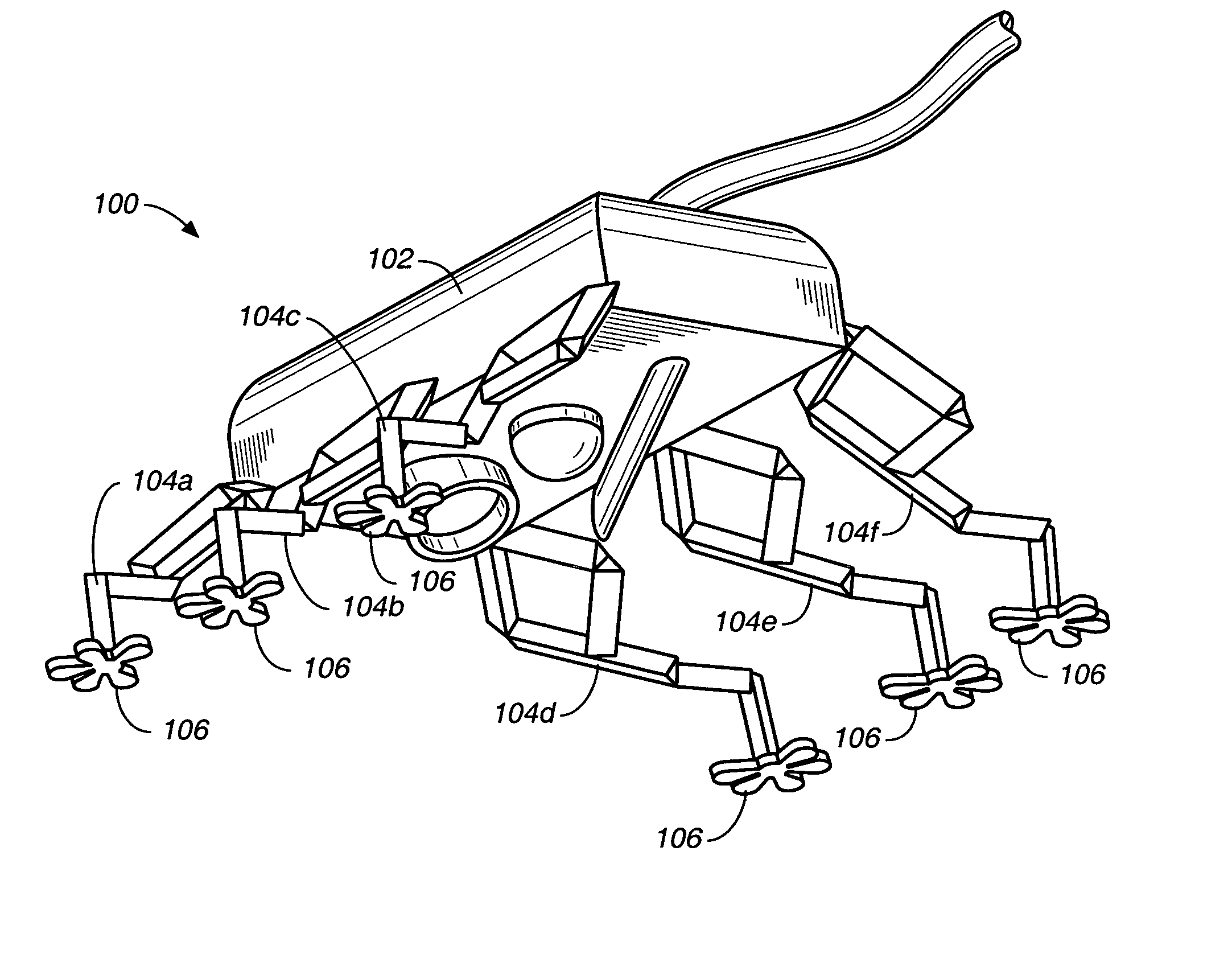
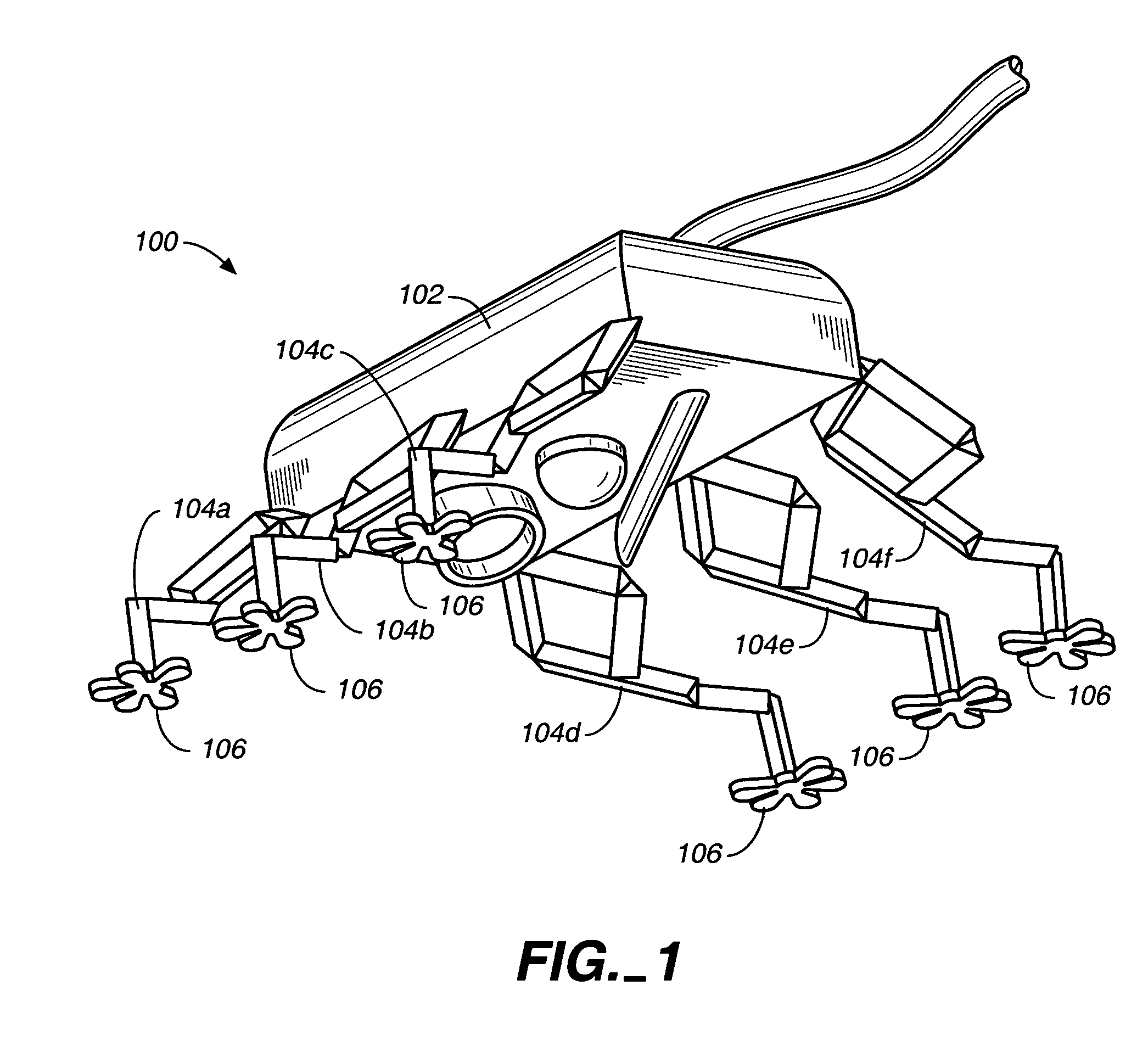
[0020] As further detailed herein, surgical instruments capable of adhering to organs and other tissues during minimally invasive surgery are provided. The surgical instruments include a micromechanical structure that adheres to tissues by micro-fibers via van der Waal's interactions. The micromechanical structure capable of adhering and moving along the surface of tissues, including moving tissues such as the heart muscle. Unlike conventional surgical robots, the surgical device can move on and in conjunction with the moving tissue such as a beating heart.

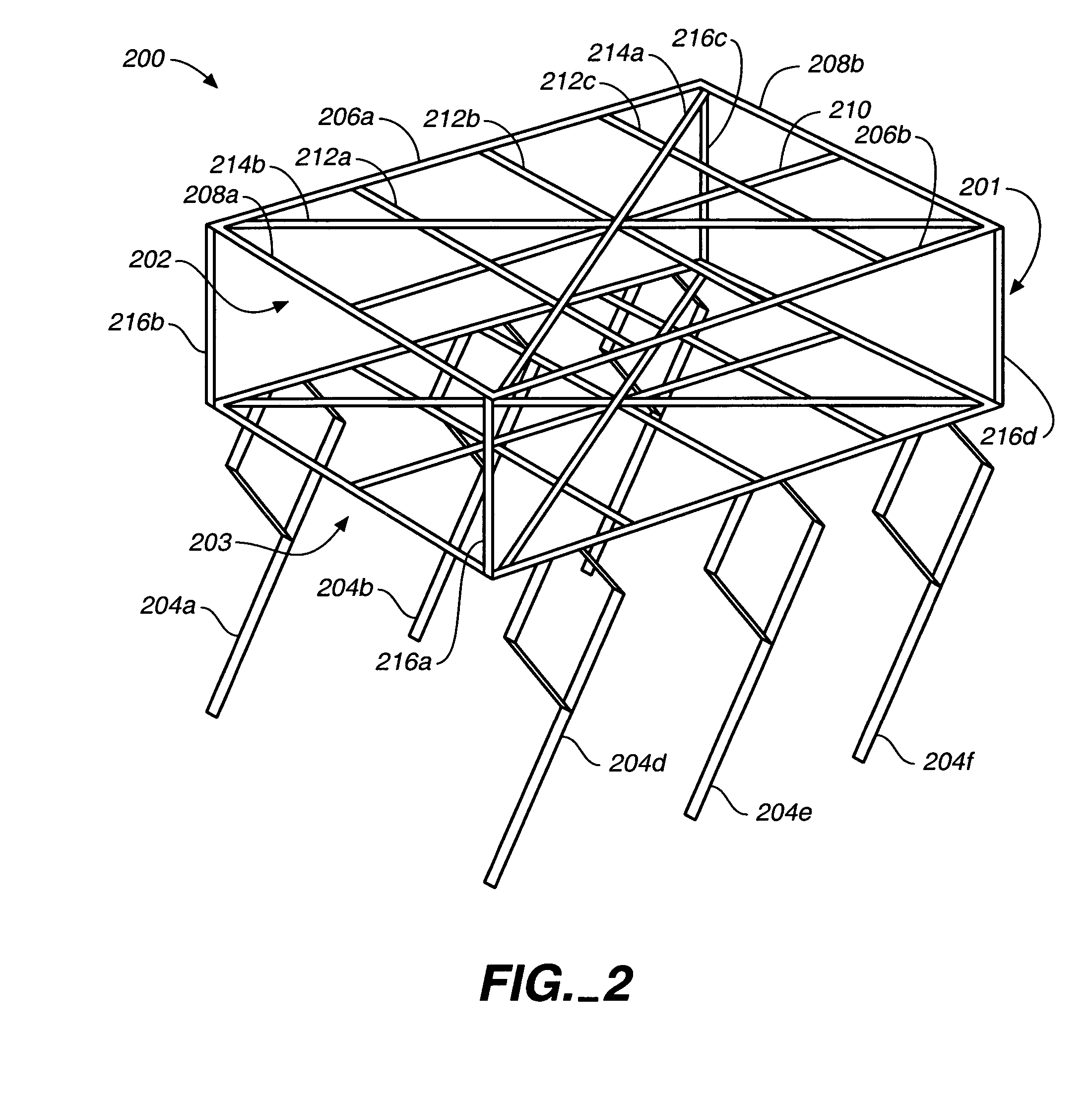
[0021] With reference t...


PUM


Abstract

A surgical device capable of adhering to tissues is disclosed herein. The surgical device includes a micromechanical frame moveably linked to a plurality of micromechanical appendages. A plurality of nano-fibers that mimic adhesion of the Tokay Gecko are disposed at the terminus of each protrusion.

Description

RELATED APPLICATIONS [0001] This application claims benefit of U.S. Provisional Application No. 60 / 508,342, filed Oct. 3, 2003, which is incorporated herein by reference in its entirety.[0002] The U.S. Government has a paid-up license in the invention and the right in limited circumstances to require the patent owner to license others on reasonable terms as provided by the terms of Grant (Contract) No. IIS 0083472 and DMII 0115091 awarded by the National Science Foundation (NSF), and N00014-98-0671 awarded by ONR MURI.BACKGROUND [0003] 1. Field [0004] This application relates generally to the fabrication and use of nano-scale adhesive structures disposed on surgical instruments. [0005] 2. Related Art [0006] One of the most difficult challenges in surgical methods is carrying out surgery as minimally invasively as possible. The ability to guide surgical instruments remotely has dramatically improved surgical methods, frequently allowing surgery to be less invasive, to improve healing...

Claims


Application Information

Patent Timeline
Patent Type & Authority Applications(United States)
IPC IPC(8): A61B10/00A61B17/00A61B17/02A61B17/068A61B17/08A61B19/00
CPCA61B10/00A61B17/00234A61B17/0218A61B17/068A61B17/085A61B19/22A61B2019/2249A61B2017/00402A61B2017/00526A61B2017/00703A61B2017/00858A61B2017/0243A61B2017/00345A61B34/70A61B34/72
Inventor SVERDUK, LEROYSAHAI, RANJANAFEARING, RONALD S.
Owner RGT UNIV OF CALIFORNIA
Why Eureka
Try Eureka