Device for enabling reduced motion-related artifacts in parallel magnetic resonance imaging

a technology of motion-related artifacts and parallel magnetic resonance imaging, which is applied in the field of medical imaging, can solve problems such as pernicious sources of errors, errors in calibration, and images with aliasing defects called artifacts or ghost artifacts, and achieve the effect of eliminating any potential coil-displacement-related reconstruction effect or artifa

Inactive Publication Date: 2005-06-23
ROBERTS TIMOTHY PAUL LESLIE +2
View PDF12 Cites 11 Cited by
  • Summary
  • Abstract
  • Description
  • Claims
  • Application Information

AI Technical Summary

Benefits of technology

[0009] In accordance with a first aspect, the present invention provides several embodiments of a device for physically separating RF imaging coils from any source of movement, thereby eliminating any potential coil-displacement related reconstruction effect or artifact. The device can be used to enable parallel imaging of the abdomen, pelvis and other moving body parts such that normal or abnormal patient movement does not displace the coil elements between the calibration scan and the imaging scans.
[0011] In another aspect of the invention there is provided a device for retrofitting to a magnetic resonance imaging apparatus for physically separating RF imaging coils from any source of movement by a patient thereby eliminating any potential coil-displacement related reconstruction effect or artifact in parallel magnetic resonance imaging, comprising:

Problems solved by technology

However, when the under-sampled k-space data is converted to an MRI image, the resulting images have aliasing defects called artifacts or ghost artifacts.
Accurate knowledge of coil sensitivities is crucial for parallel MRI, and errors in calibration represent one of the most common and the most pernicious sources of error in parallel image reconstructions.
However, in practice if the coil moves during data acquisition, for example due to breathing, the estimated coil sensitivities will be compromised, ghost artifacts will be generated and the resulting image quality will degrade.
An attractive opportunity for parallel imaging exists in the abdomen and pelvis, where scans are typically limited in quality by the requirement for acquisition to be completed during a single period of suspended respiration (breath-hold).
Accordingly, the problem of varying coil sensitivity is particularly pronounced for imaging of the abdomen, where calibration scans and images are typically acquired during separate periods of suspended respiration (breath-holds) which are rarely precisely reproducible.
In fact, the problem is so severe that the ghost artifact mechanism may impose a limitation on the use of these accelerated imaging methods in some settings.
However, these approaches are limited by patient compliance and reproducibility.
Other applications of MR imaging such as “interventional” or study of joint kinematics involve other types of motion and hence cannot use an approach related to minimizing coil movement due to respiration.

Method used


Image

  • Device for enabling reduced motion-related artifacts in parallel magnetic resonance imaging
  • Device for enabling reduced motion-related artifacts in parallel magnetic resonance imaging
  • Device for enabling reduced motion-related artifacts in parallel magnetic resonance imaging

Examples


Embodiment Construction

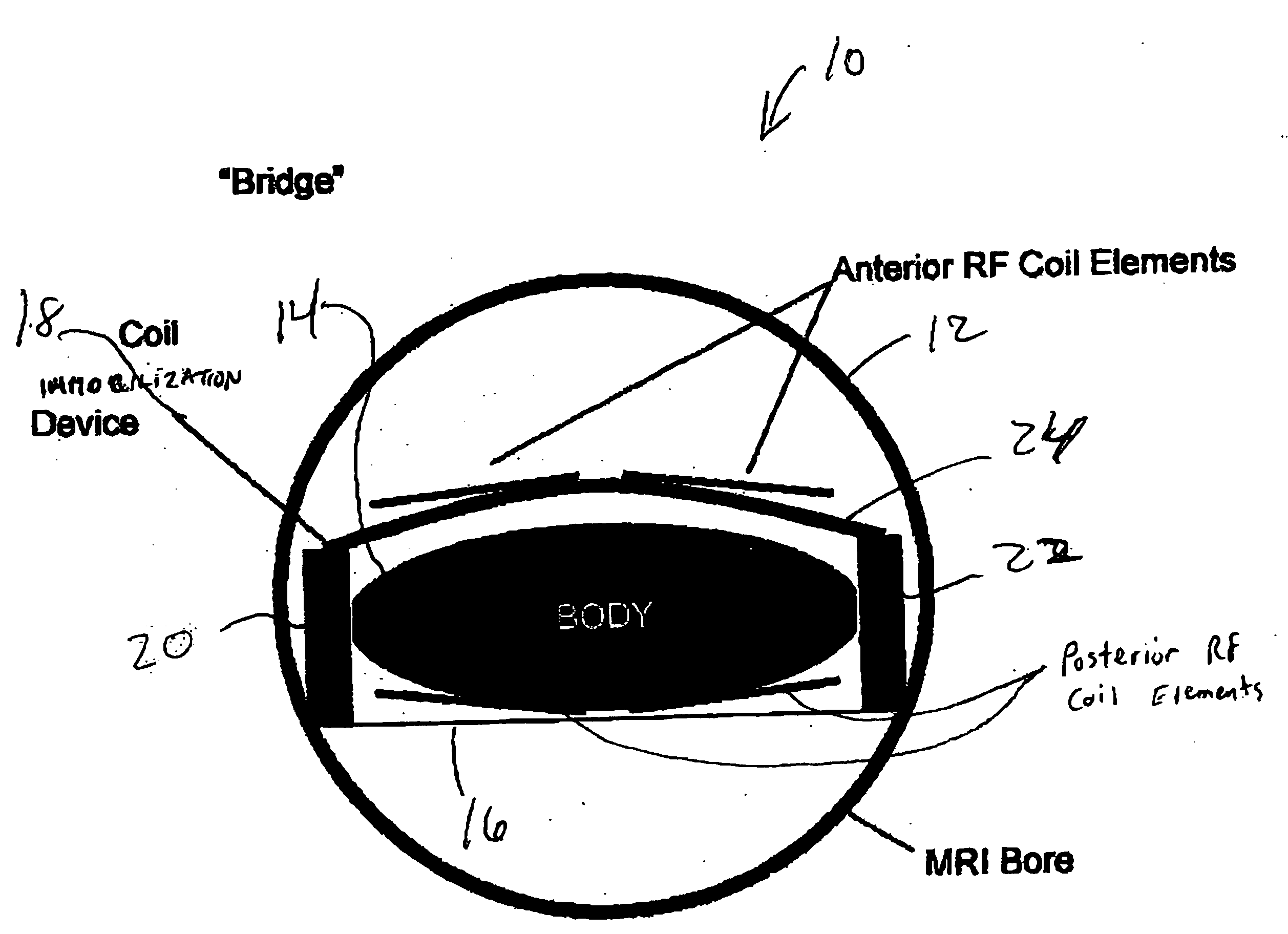
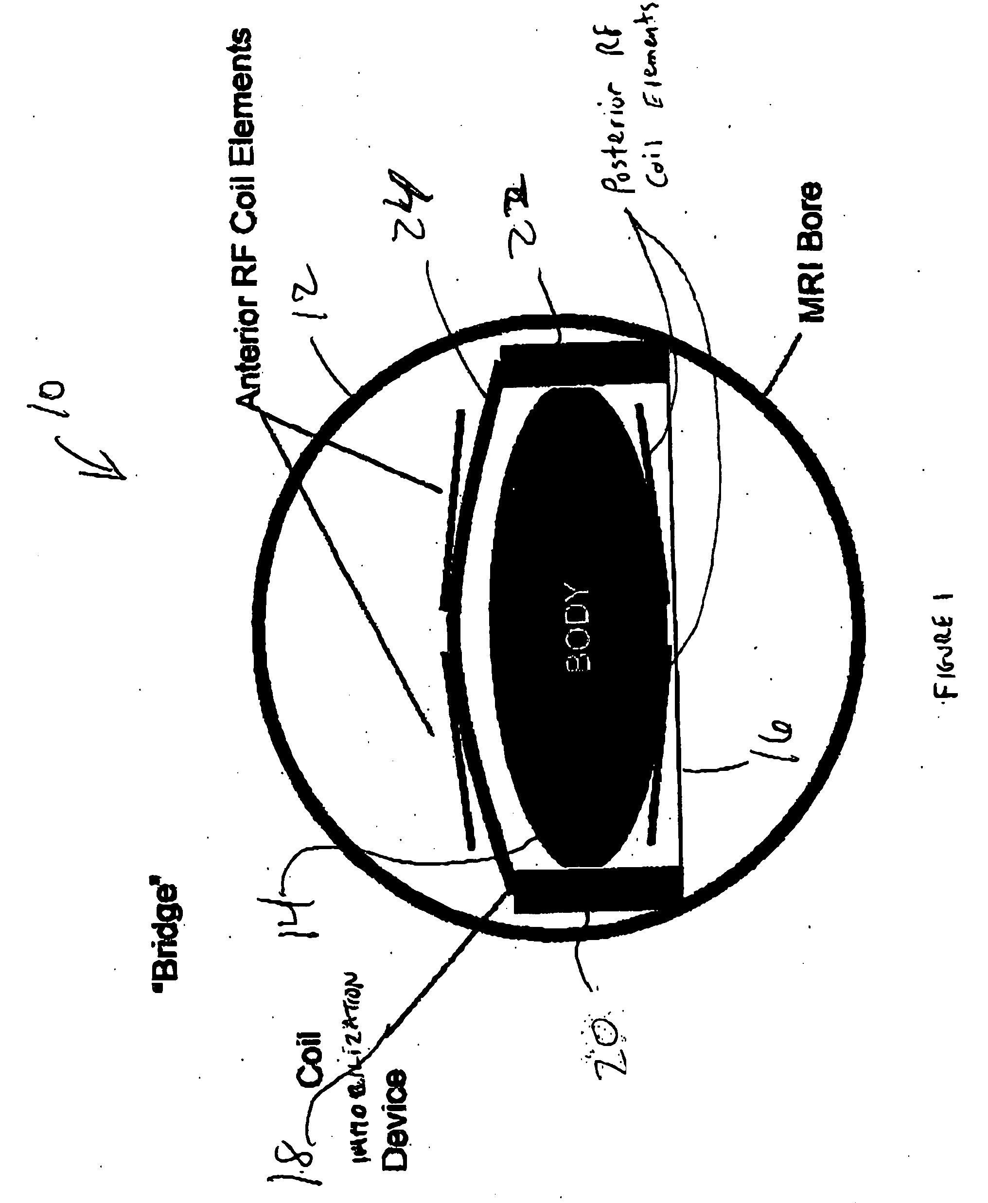
[0024] Referring now to FIG. 1, shown therein is a block diagram of an embodiment of a coil immobilization device in accordance with the present invention. FIG. 1 is a cross-sectional view of the body of a patient in the MRI bore of an MRI scanner. The MRI scanner includes excitation RF coils (not shown) for generating excitation magnetic fields that create changes in the magnetic spin moments of the atoms in the patient's body. The changes in the magnetic spin moments provides data that is recorded by the anterior and posterior imaging RF coil elements.

[0025] The anterior RF coil elements are statically held in place by the coil immobilization device. The posterior RF coil elements are integrated into a cushion (not shown) or the platform upon which the patient lies. The posterior RF coils cannot move regardless of whether the patient moves.

[0026] In current practice by those skilled in the art, the anterior RF coils are placed directly on the outer wall of the patient's body for...


PUM


Abstract

The present invention provides several embodiments of a device for physically separating RF imaging coils from any source of movement thereby minimizing potential coil-displacement related reconstruction effect or artifact. The device can be used to enable parallel imaging of the abdomen, pelvis and other moving body parts such that normal or abnormal patient movement does not displace the coil elements between the calibration scan and the subsequent imaging scans.

Description

CROSS REFERENCE TO RELATED U.S APPLICATION [0001] This patent application relates to, and claims the priority benefit from, U.S. Provisional Patent Application Ser. No. 60 / 525,832 filed on Dec. 1, 2003, which is incorporated herein by reference in its entirety.FIELD OF THE INVENTION [0002] The present invention relates to a method and device used in medical imaging. In particular, the invention is related to a method and device for enabling reduced motion-related displacement artifacts in parallel magnetic resonance imaging. BACKGROUND OF THE INVENTION [0003] Magnetic Resonance Imaging (MRI) is based on the absorption and emission of energy in the radio frequency range. A patient is placed in a magnetic resonance scanner that provides a uniform magnetic field that causes the alignment of the moments of the magnetic spin of atoms contained within the patient. The magnetic resonance scanner further provides multiple coils that apply a transverse magnetic field, generated by RF pulses,...

Claims


Application Information

Patent Timeline
Patent Type & Authority Applications(United States)
IPC IPC(8): A61B5/055G01R33/20G01R33/3415G01R33/561
CPCG01R33/5611G01R33/3415
Inventor ROBERTS, TIMOTHY PAUL LESLIESUSSMAN, MARSHALL STEPHENFLAGG, ELISSA JILL
Owner ROBERTS TIMOTHY PAUL LESLIE
Why Eureka
Try Eureka