Image reporting method and system

a reporting method and image technology, applied in the field of image reporting methods and systems, can solve the problems of lack of standardization, consistency, accountability, efficiency, and lack of standardized methods in the way in which reports are generated, and achieve the effects of efficient and systematic generation of radiology reports, facilitating data entry into searchable databases, and accelerating hospital billing and collection

Inactive Publication Date: 2005-07-07
WAKE FOREST UNIV HEALTH SCI INC
View PDF46 Cites 70 Cited by
  • Summary
  • Abstract
  • Description
  • Claims
  • Application Information

AI Technical Summary

Benefits of technology

[0008] The present invention relates to a new reporting method and system for reporting the findings of an expert's analysis of image data and, more specifically, to a computer system and computer-implemented method for reporting an expert's findings relative to an analysis of image data. The method and system are based on a new reporting paradigm. The paradigm forms the basis of a radiology practice management system that can efficiently and systematically generate radiology reports, facilitate data entry into searchable databases, support clinical trials and outcomes analyses, and expedite hospital billing and collections. One fundamental aspect of this paradigm is that an expert, e.g. a radiologist, identifies a diagnostically significant feature on an image and attaches a location:description code, or in the case of radiology an anatomical:pathological code, to the location of that feature in order to create a finding, or in the case of radiology a diagnostic finding. The anatomical:pathological code includes the anatomical location followed by a pathological description. Optionally, further attributes of that finding, such as dimensional measurements (e.g., length, area, and volume), audio descriptions, 3D rendered snapshots, etc., may be automatically appended to the diagnostic finding as secondary attributes of the diagnostic finding. All of this information is automatically captured in an intuitive workflow scheme transparent to the expert, and stored in a database. The expert may continue to identify additional diagnostically significant features and create diagnostic findings in any order. At the end of the expert's evaluation of the image(s), the system sorts the diagnostic findings by selected or predetermined categories. In a medical field, these predetermined categories may be anatomical categories. The diagnostic findings are further prioritized by the severity of the diagnosis in order to alert the report recipient, e.g., a clinician. The expert can edit and approve a multimedia report, which may be delivered to an Internet server for immediate access, sent to a database, sent by automated voice, fax or e-mail to the clinician, or any combination thereof. The radiologist can sign the report by electronic or voice signature. The final report presentation may be further customized to satisfy the needs of the clinician.

Problems solved by technology

Image reporting as currently practiced suffers from a lack of standardization, consistency, accountability, and efficiency.
A root cause of these problems is the manner in which reports are generated, beginning with the lack of a standardized report format, particularly in the medical field of radiology.
This inconsistency among the radiologists' reporting styles often hinders the correlation of the reported findings with the actual images by the recipients of the reports.
Variability in the reporting styles also impedes on-going monitoring of specific findings from different examinations on the same patient, a task that is critical for patient care and time-consuming for radiologists.
Further, traditional radiology reporting practices do not support data mining, a powerful tool which is useful in clinical trials, epidemiology studies, and outcomes analyses.
In addition, conventional reporting practices often provide no mechanism to allow the radiologist to account for the effective communication of critical report information to the recipient.
To the contrary, however, radiologists are often held accountable for ensuring that proper action is taken on significant findings and are held liable for malpractice when proper action is not taken.
A major complaint of such clinicians against radiologists and their reporting practices involves point of service.

Method used


Image

  • Image reporting method and system
  • Image reporting method and system
  • Image reporting method and system

Examples


Embodiment Construction

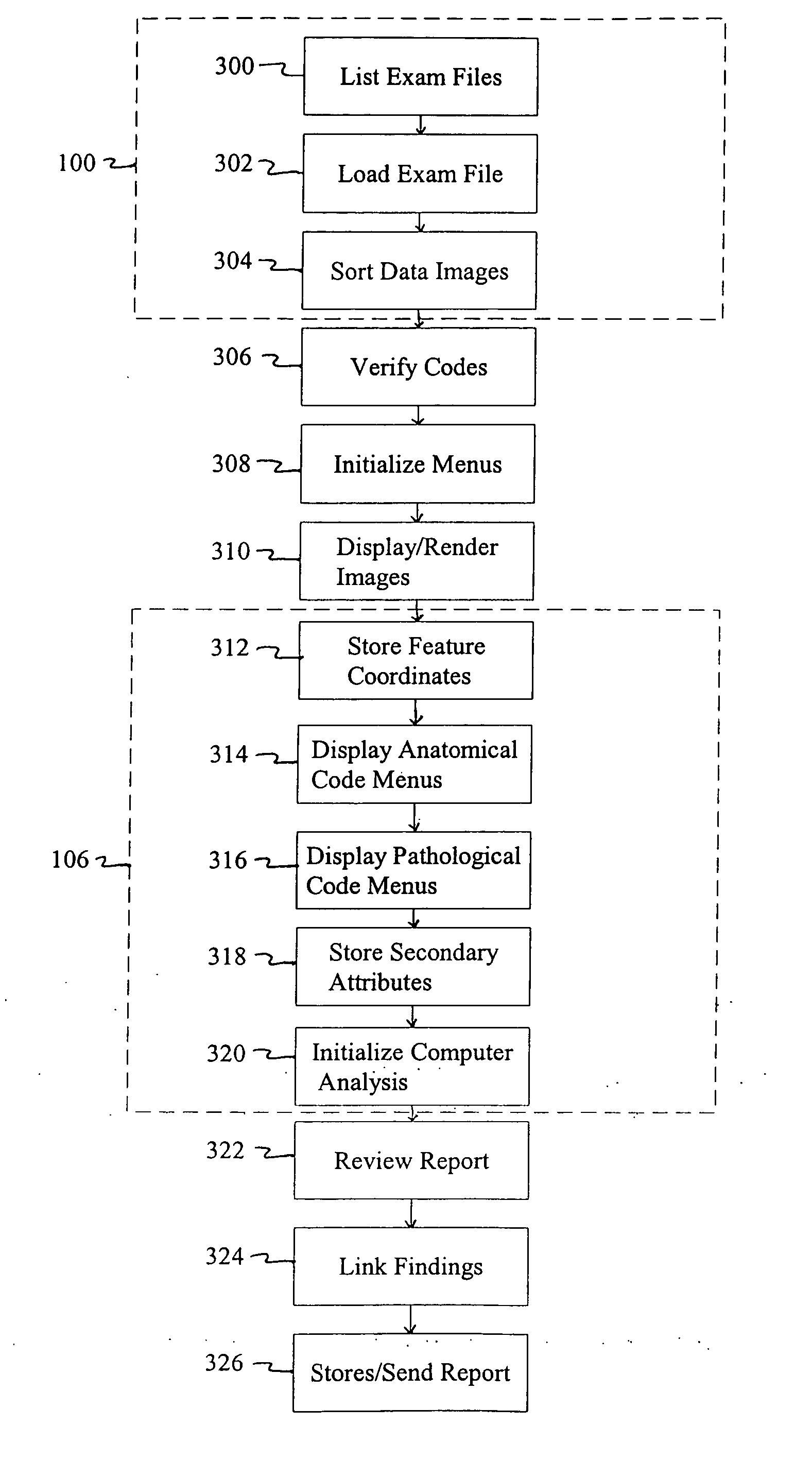
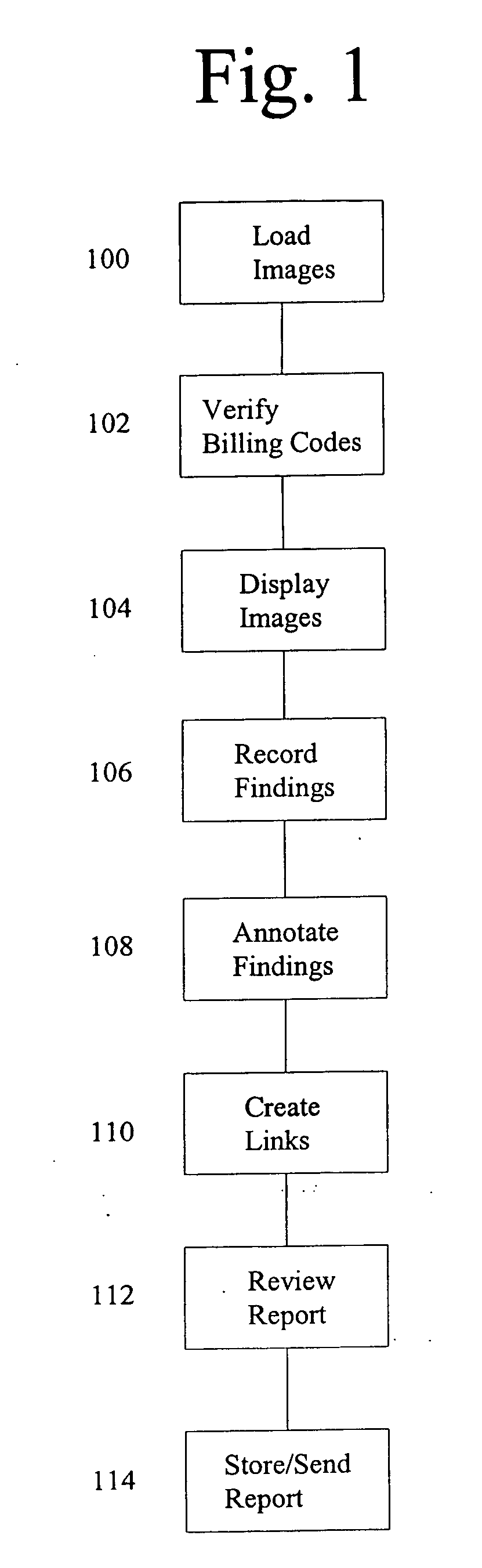
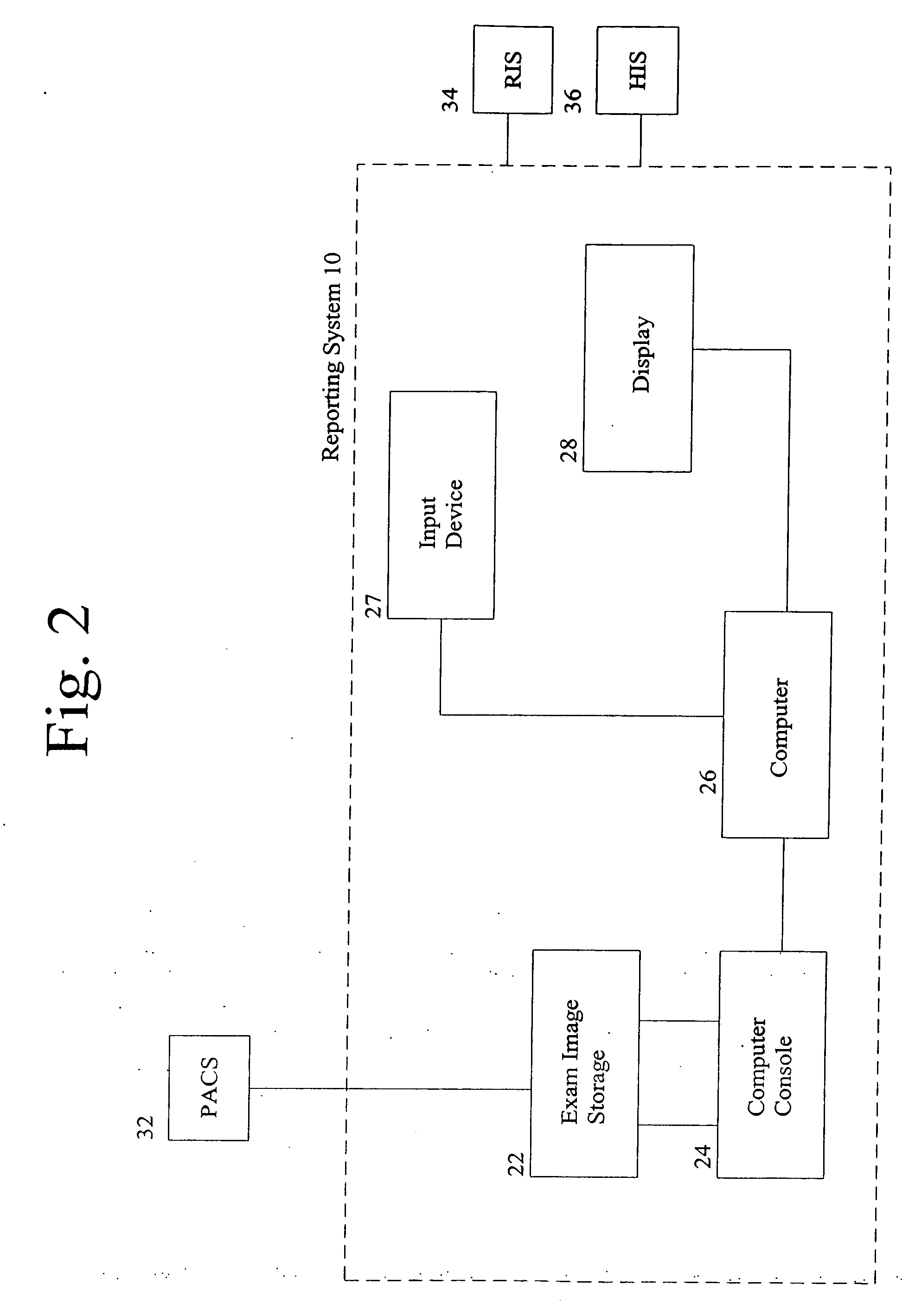
[0018] A method and system are provided for generating and communicating reports containing an expert's analysis of image data as generally depicted in FIGS. 1 and 2. In addition, a computer-implemented method and a computer system function to create a database of the expert's findings from which a report is generated and from which data mining and other analyses may be conducted. The database can be a computer searchable database and may be a relational computer database.

[0019] The method and system of the present invention are applicable to any field which relates to an expert's analysis of images. In particular, however, the method and system of the present invention are well-suited to image analysis found in medical applications. As such, the method and system of the present invention are illustrated in the accompanying figures and description in terms of the medical field of radiology.

[0020] The method and system are particularly well-suited to the analysis of digital images....


PUM

PropertyMeasurementUnit
opacityaaaaaaaaaa
volumeaaaaaaaaaa
surface renderingaaaaaaaaaa

Abstract

A method and system are provided to report the findings of an expert's analysis of image data. The method and system are based on a reporting system that forms the basis of an image management system that can efficiently and systematically generate image reports, facilitate data entry into searchable databases for data mining, and expedite billing and collections for the expert's services. The expert identifies a significant finding on an image and attaches a location:description code to the location of that finding in order to create a significant finding and an entry into a database. Further descriptions of that finding, such as dimensional measurements, audio descriptions, 3D rendered snapshots, etc., may be automatically appended to the finding as secondary attributes of the finding within the database. At the end of the expert's evaluation of the image(s), the system sorts the findings in the database and presents the findings by prioritized categories. The expert edits and approves a multimedia report, which may be delivered to an Internet server for immediate access, archived in the database, sent by automated voice, fax or e-mail to an end-user, or any combination thereof.

Description

[0001] Applicants claim the benefit of priority to U.S. Provisional Application 60 / 147,914, filed on Aug. 9, 1999, the contents of which are incorporated herein by reference.FIELD OF THE INVENTION [0002] The present invention relates generally to an image reporting method and system and more particularly to a method and computer-implemented procedure for creating electronic, multimedia reports based on a new reporting paradigm. BACKGROUND OF THE INVENTION [0003] Image reporting as currently practiced suffers from a lack of standardization, consistency, accountability, and efficiency. A root cause of these problems is the manner in which reports are generated, beginning with the lack of a standardized report format, particularly in the medical field of radiology. [0004] Radiologists generally review images of a body structure and dictate narrative descriptions of their image findings followed by summary statements. Transcriptionists then transcribe the dictated statements and either ...

Claims


Application Information

Patent Timeline
Patent Type & Authority Applications(United States)
IPC IPC(8): G06F17/30G06Q10/00A61B5/00G06T7/00G11B20/12G16H15/00G16H30/40
CPCG06F17/30056Y10S707/99948G06F19/321G06F19/3406G06F19/3425G06F19/345G06F19/3487G06Q10/10G06Q30/04G06Q50/24G06T7/0012G11B2020/10944Y10S707/99945Y10S128/922G06F17/30256G16H80/00G16H40/63G16H50/20G16H15/00G06F16/5838G06F16/4393G16H30/40
Inventor VINING, DAVID J.GE, YAORONGAHN, DAVID K.STELTS, DAVID R.
Owner WAKE FOREST UNIV HEALTH SCI INC
Why Eureka
Try Eureka