Electrode sleeve for biopsy device

a biopsy device and electrode sleeve technology, applied in medical science, surgery, vaccination/ovulation diagnostics, etc., can solve the problem that no single procedure is ideal for all cases, and achieve the effect of controlling cautery

Inactive Publication Date: 2005-09-15
DEVICOR MEDICAL PROD
View PDF20 Cites 34 Cited by
  • Summary
  • Abstract
  • Description
  • Claims
  • Application Information

AI Technical Summary

Benefits of technology

[0011] In one embodiment, the present invention provides a pair of electrodes attached to the exterior of a sleeve. The electrode sleeve can be used with the handheld biopsy device, such as a MAMMOTOME® biopsy instrument. The sleeve can be sized to slide over the piercer of the biopsy device and the sleeve can have a window sized to match the port on the piercer. The pair of electrodes can be disposed in association with the window, with one electrode extending along each side of the window. A gap between the electrodes allows treatment of tissue between the electrode gap with radio frequency (RF) energy to coagulate and create hemostasis. The knife tip of the handheld biopsy device can act as a third electrode, allowing treatment between each of the side electrodes and the knife tip structure. The sleeve uses an interface board to systematically control cautery by switching from one electrode to the other, or from one electrode to the knife tip.

Problems solved by technology

The type of biopsy used depends mainly on various factors present in the patient, and no single procedure is ideal for all cases.
Bleeding of tissue at the knife tip and around the piercer of the biopsy device may occur with current biopsy devices.

Method used


Image

  • Electrode sleeve for biopsy device
  • Electrode sleeve for biopsy device
  • Electrode sleeve for biopsy device

Examples


Embodiment Construction

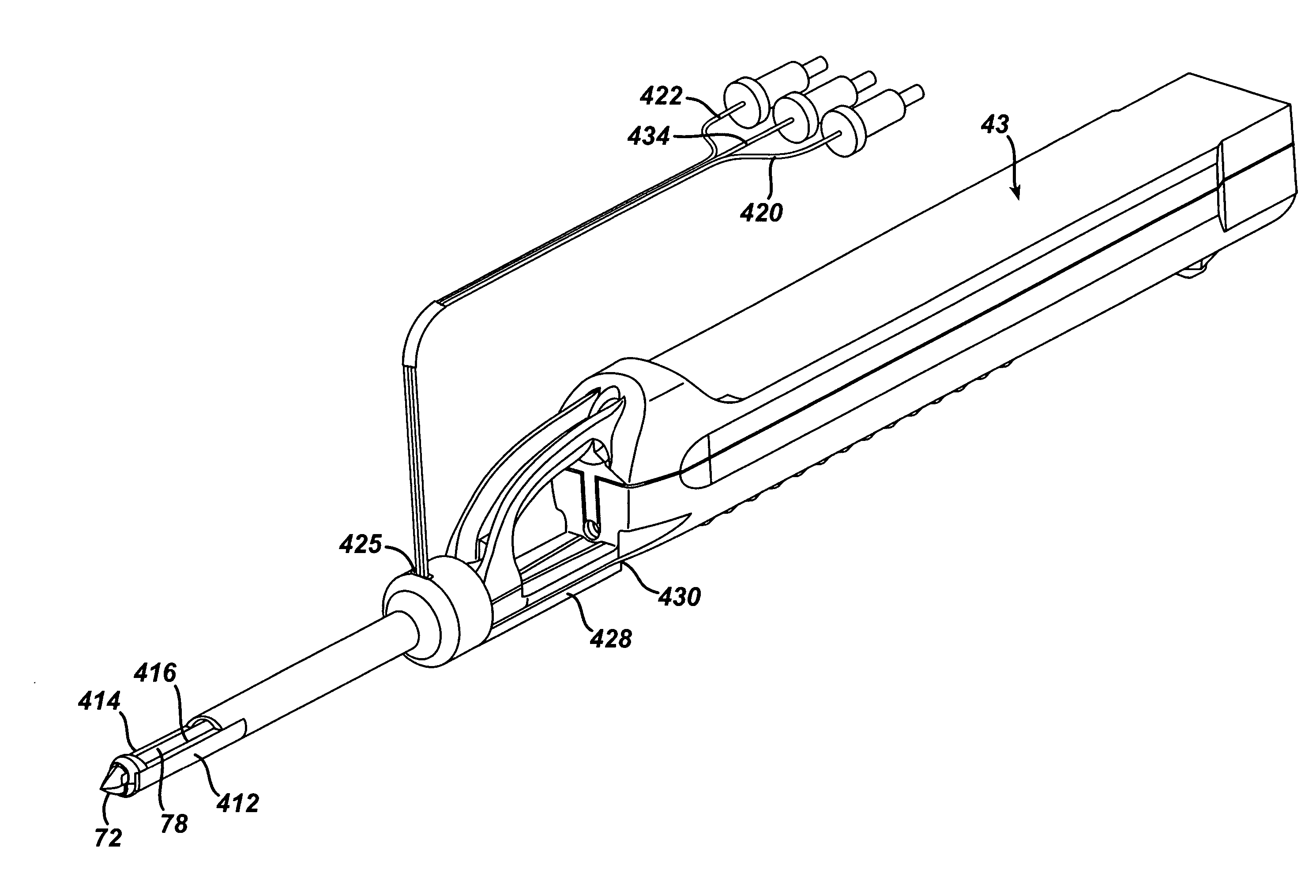
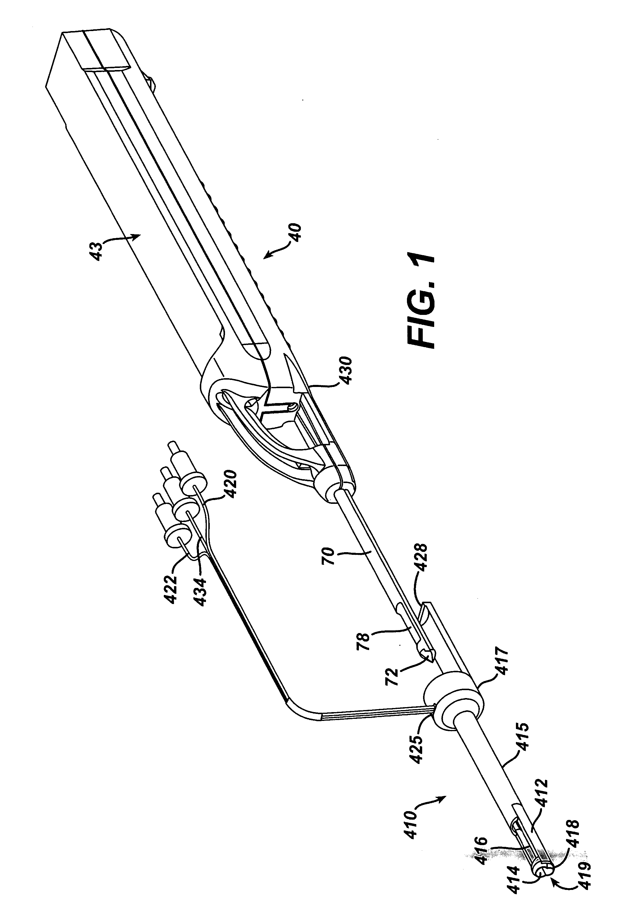
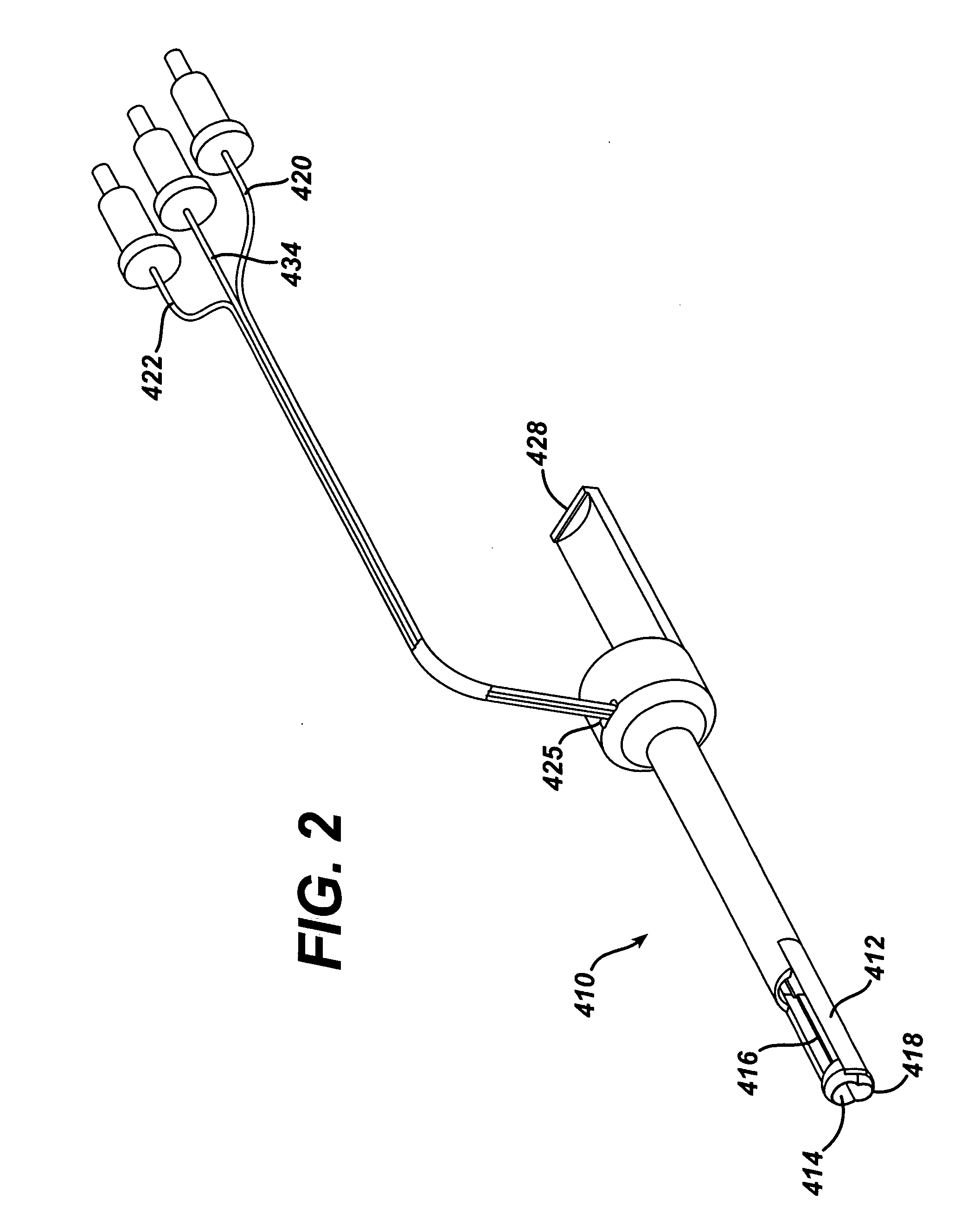
[0027] Referring now to the Figures, in which like numerals indicate like elements, FIG. 1 discloses an exploded view of a probe assembly 40 and a sleeve 410. Probe assembly 40 may be a probe assembly as disclosed in U.S. Pat. No. 6,273,862, “Surgical Device for the Collection of Soft Tissue,” the entire contents of which are hereby incorporated herein by reference. A suitable probe assembly 40 is a part of the MAMMOTOME® breast biopsy instrument, available from Ethicon Endo-Surgery, Cincinnati, Ohio. Probe assembly 40 can include a hollow cannular piercer 70 extending distally from a hollow handle 43, a distal piercing knife tip 72 at the distal end of piercer 70, and a groove 430 on the underside of handle 43. Sleeve 410 can include a first electrode 412, a second electrode 414, a tissue receiving window 416, an electrode gap 418, a connector 428, a first wire 420 attached to electrode 412, a second wire 422 attached to electrode 414, and a third wire 434. Sleeve 410 is sized to s...


PUM


Abstract

A handheld biopsy device is provided for the collection of soft tissue samples from a surgical patient. At least one electrode is provided, such as between a handle and the knife tip of the biopsy device. The electrodes can be provided on a sleeve. The sleeve with electrodes can be releasably supported on the biopsy device.

Description

FIELD OF THE INVENTION [0001] The present invention is related generally to devices and methods for the collection of soft tissue. More particularly, it relates to a device and a method to improve coagulation of tissue with biopsy devices used for the collection of soft tissue. BACKGROUND OF THE INVENTION [0002] The diagnosis and treatment of patients with cancerous tumors, pre-malignant conditions, and other disorders has long been an area of intense investigation. Non-invasive methods for examining tissue include palpation, X-ray, MRI, CT, and ultrasound imaging. When the physician suspects that a tissue may contain cancerous cells, a biopsy may be done using either an open procedure or a percutaneous procedure. For an open procedure, a scalpel is used by the surgeon to create a large incision in the tissue in order to provide direct viewing and access to the tissue mass of interest. The entire mass (excisional biopsy) or a part of the mass (incisional biopsy) may then be removed....

Claims


Application Information

Patent Timeline
Patent Type & Authority Applications(United States)
IPC IPC(8): A61B10/02A61B10/00A61B18/12A61B18/14
CPCA61B10/0275A61B2010/0208A61B18/14
Inventor VOEGELE, JAMES W.
Owner DEVICOR MEDICAL PROD
Why Eureka
Try Eureka