System and method for visualization and navigation of three-dimensional medical images

a three-dimensional medical image and system technology, applied in the field of system and method for visualization and navigation of three-dimensional medical images, can solve the problems of difficult diagnosis of stenosis or other abnormalities, lack of efficient or intuitive means to navigate through virtual organs, etc., and achieve the effect of enabling user interaction

Inactive Publication Date: 2005-10-13
VIATRONIX
View PDF0 Cites 289 Cited by
  • Summary
  • Abstract
  • Description
  • Claims
  • Application Information

AI Technical Summary

Problems solved by technology

However, while stacks of such slices may be useful for analysis, they do not provide an efficient or intuitive means to navigate through a virtual organ, especially one as tortuous and complex as the colon, or arteries.
For example, when imaging blood vessels, cross-sections merely show slices through vessels, making it difficult to diagnose stenosis or other abnormalities.

Method used


Image

  • System and method for visualization and navigation of three-dimensional medical images
  • System and method for visualization and navigation of three-dimensional medical images
  • System and method for visualization and navigation of three-dimensional medical images

Examples


Embodiment Construction

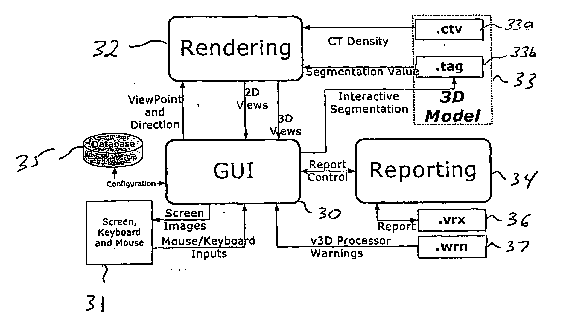
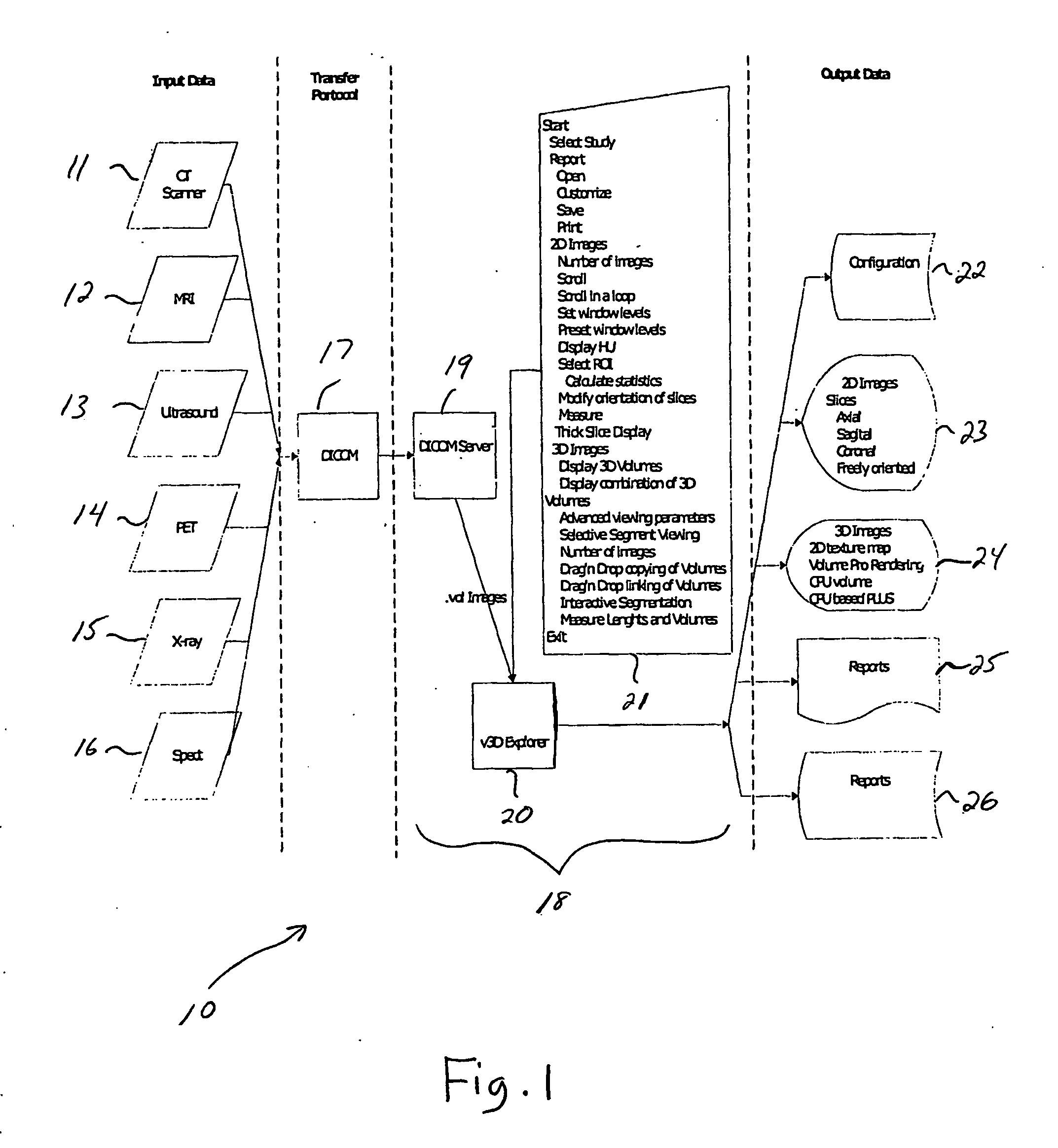
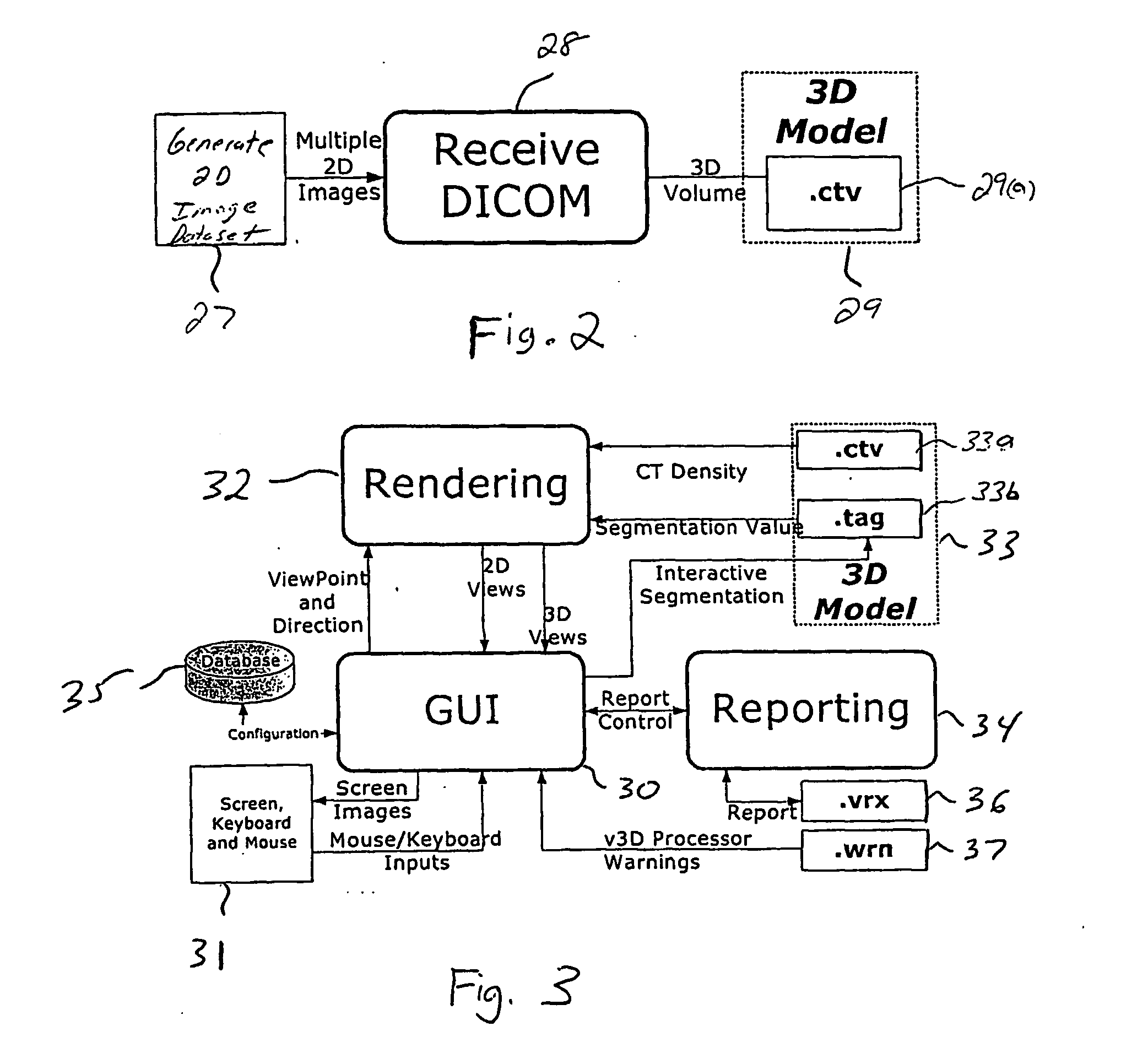
[0030] The present invention is directed to medical imaging systems and methods for assisting in medical diagnosis and evaluation of a patient. Imaging systems and methods according to preferred embodiments of the invention enable visualization and navigation of complex 2D and 3D models of internal organs, and other components, which are generated from 2D image datasets generated by a medical imaging acquisition device (e.g., MRI, CT, etc.).

[0031] It is to be understood that the systems and methods described herein in accordance with the present invention may be implemented in various forms of hardware, software, firmware, special purpose processors, or a combination thereof. Preferably, the present invention is implemented in software as an application comprising program instructions that are tangibly embodied on one or more program storage devices (e.g., magnetic floppy disk, RAM, CD Rom, ROM and flash memory), and executable by any device or machine comprising suitable architect...


PUM


Abstract

A user interface (90) comprises an image area that is divided into a plurality of views for viewing corresponding 2-dimensional and 3-dimensional images of an anatomical region. Tool control panes (95-101) can be simultaneously opened and accessible. The segmentation pane (98) enables automatic segmentation of components of a displayed image within a user-specified intensity range or based on a predetermined intensity

Description

CROSS-REFERENCE TO RELATED APPLICATION [0001] This application claims priority to U.S. Provisional Application No. 60 / 331,799, filed on Nov. 21, 2001, which is fully incorporated herein by reference.COPYRIGHT NOTICE [0002] A portion of the disclosure of this patent document contains material which is subject to copyright protection. The copyright owner has no objection to the facsimile reproduction by any one of the patent document or the patent disclosure, as it appears in the patent and Trademark Office patent file or records, but otherwise reserves all copyright rights whatsoever. TECHNICAL FIELD OF THE INVENTION [0003] The present invention relates generally to systems and methods for aiding in medical diagnosis and evaluation of internal organs (e.g., colon, heart, etc.) More specifically, the invention relates to a 3D visualization (v3D) system and method for assisting in medical diagnosis and evaluation of internal organs by enabling visualization and navigation of complex 2D...

Claims


Application Information

Patent Timeline
Patent Type & Authority Applications(United States)
IPC IPC(8): A61B6/03A61B17/00A61B17/22A61B19/00
CPCA61B5/02007A61B6/5247A61B6/037A61B6/463A61B6/503A61B6/504A61B6/5235A61B8/468A61B8/5238A61B17/22A61B19/5244A61B19/56A61B2017/00243A61B2019/505A61B2019/5291A61B2019/564A61B2019/566G01S7/52073G01S7/52074G01S7/52084G01S15/8993A61B6/032A61B34/20A61B34/25A61B2034/105A61B2034/254A61B2034/256A61B2090/365
Inventor BITTER, INGMARLI, WEIMEISSNER, MICHAELDACHILLE, FRANK C.GRIMM, SORENECONOMOS, GEORGE
Owner VIATRONIX
Why Eureka
Try Eureka