Method and apparatus for medical ultrasound navigation user interface

a navigation user interface and ultrasound technology, applied in the field of diagnostic ultrasound methods and systems, can solve the problems of inability of ultrasound methods and systems to acquire volumetric ultrasound data

Active Publication Date: 2005-12-22
GENERAL ELECTRIC CO
View PDF22 Cites 45 Cited by
  • Summary
  • Abstract
  • Description
  • Claims
  • Application Information

AI Technical Summary

Benefits of technology

[0005] A need exists for improved methods and systems that provide real time navigation in ultrasound volumetric data.
[0006] One embodiment, an ultrasound system is provided that includes a probe having a 2D array of transducers for acquiring ultrasound information along a plurality of scan lines through an object in real time. The scan lines are arranged to define volumetric data corresponding to a volume of interest (VOI) in a subject or patient. One such VOI may include the human heart or some portion of the human heart. The system includes a beamformer configured with scan parameter values that define the scan lines. An image buffer stores multiple data volumes acquired over time that are successively retrieved and processed by a display processor. The display processor accesses the data volumes stored in the image buffer successively to control generation of at least one of 2D and 3D renderings based on display parameters, wherein the display processor obtains from the image buffer a first data volume defined based on first scan parameter values, while the probe acquires ultrasound information for a second data volume that is entered into the image buffer. The second data volume is defined based on second scan parameter values. A navigation view presents in real time the renderings generated by the display processor with their corresponding 3...

Problems solved by technology

Heretofore, ultrasound methods and systems were unable to acquire volumetric ultrasound data at the s...

Method used


Image

  • Method and apparatus for medical ultrasound navigation user interface
  • Method and apparatus for medical ultrasound navigation user interface
  • Method and apparatus for medical ultrasound navigation user interface

Examples


Embodiment Construction

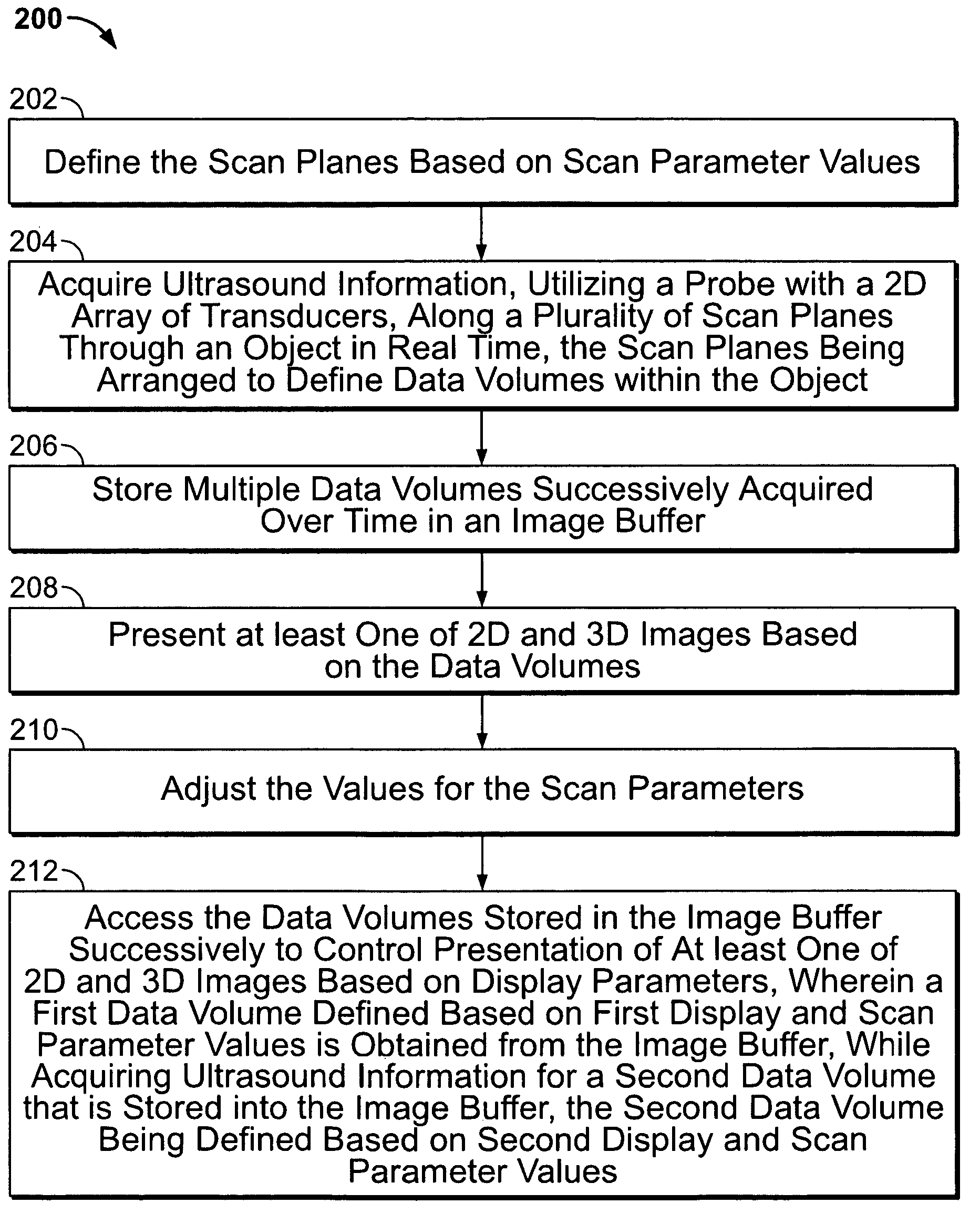
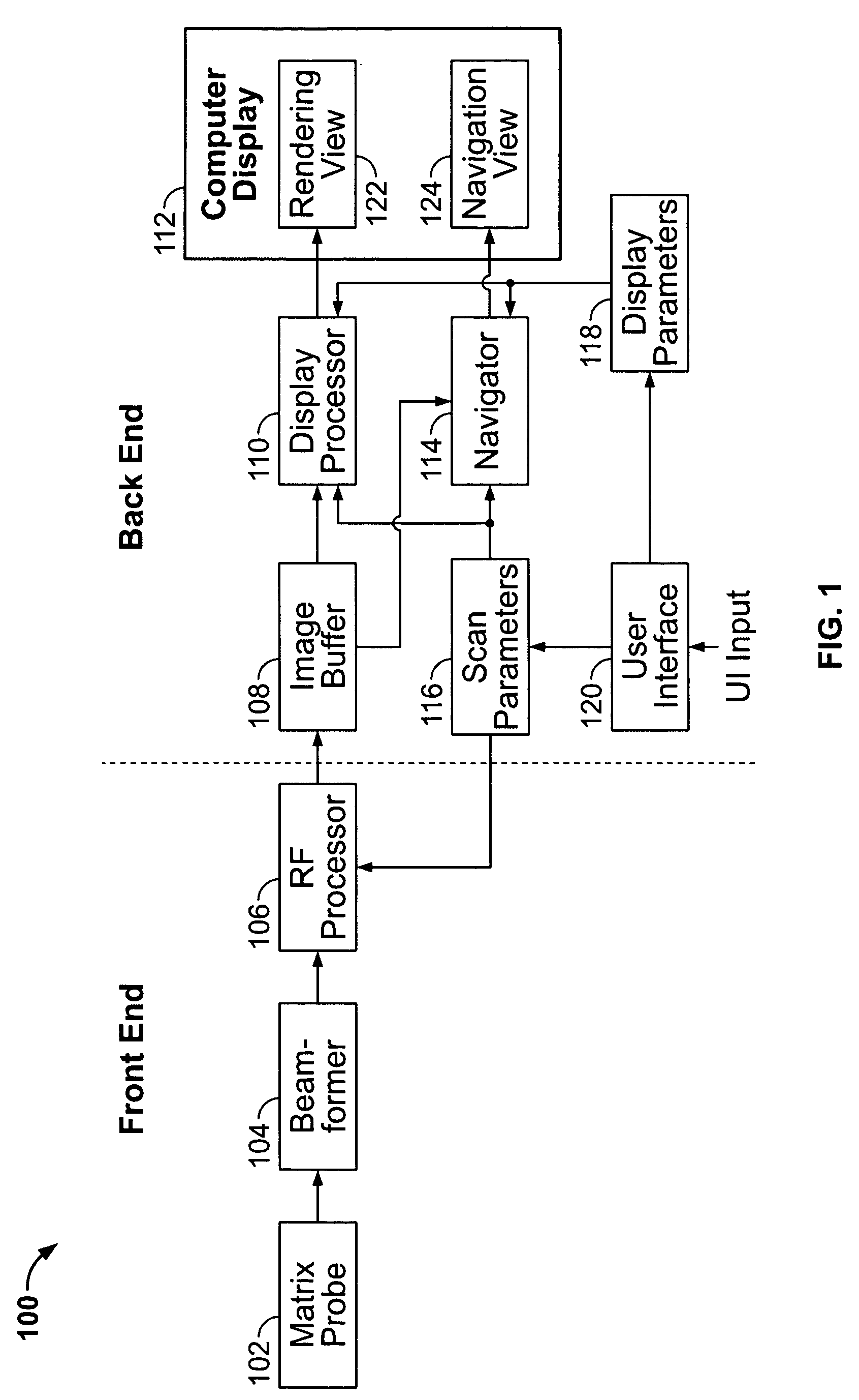
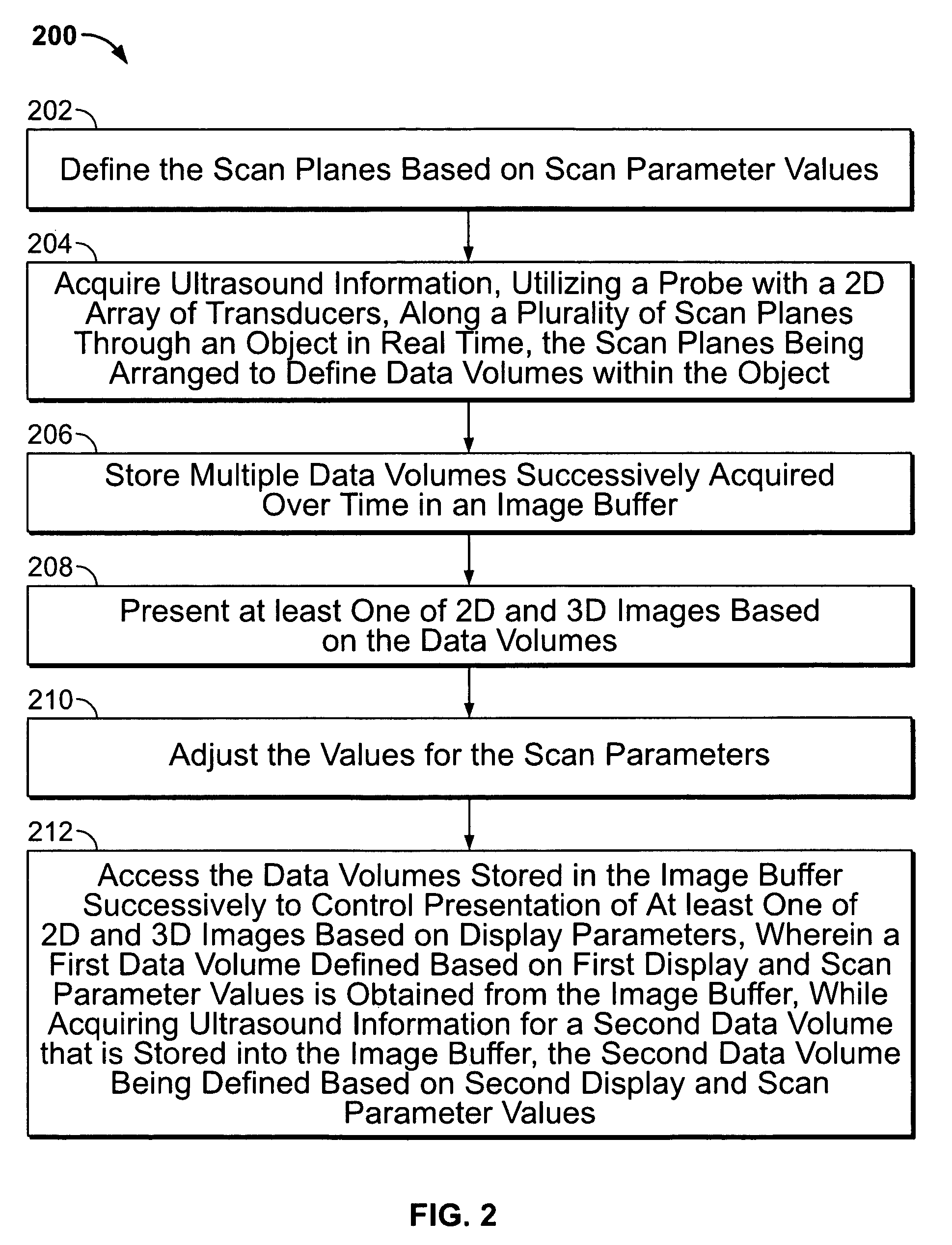
[0017]FIG. 1 is a block diagram of an ultrasound system 100 formed in accordance with an embodiment of the present invention. The ultrasound system 100 is configurable to acquire volumetric data corresponding to a volume of interest (VOI) in a subject or patient. One such VOI may include the human heart or some portion of the human heart. The ultrasound system 100 is configurable to acquire 3-dimensional (3D) volumes of data, each volume defined by an azimuth angle and elevation angle. The ultrasound system 100 includes a 2-dimensional (2D) array / matrix probe 102 that under the guidance of a beamformer 104 scans the VOI and acquires volumes of data at a rate of 15-30 volumes / sec, depending on the size of the volume (azimuth angle and elevation angle). The probe 102 receives backscattered echoes from the scanned object within the VOI and generates electrical receive signals that are combined in the beamformer 104 to form each beam / line within each scan plan. Multiple scan plans (diff...


PUM


Abstract

In one embodiment, an ultrasound system is provided that includes a probe having a 2D array of transducers for acquiring ultrasound information along a plurality of scan lines through an object in real time. The scan lines are arranged to define volumetric data corresponding to a volume of interest (VOI) in a subject or patient. One such VOI may include the human heart or some portion of the human heart. The system includes a beamformer configured with scan parameter values that define the scan lines. An image buffer stores multiple data volumes acquired over time that are successively retrieved and processed by a display processor. The display processor accesses the data volumes stored in the image buffer successively to control generation of at least one of 2D and 3D renderings based on display parameters, wherein the display processor obtains from the image buffer a first data volume defined based on first scan parameter values, while the probe acquires ultrasound information for a second data volume that is entered into the image buffer. The second data volume is defined based on second scan parameter values. A navigation view presents in real time the renderings generated by the display processor with their corresponding 3D orientation. A navigator is provided that controls the display of the navigation view in real time such that, as the user adjusts a display parameter value to change a view plane, images presented in the navigation view are updated to reflect the view plane. A user interface is provided for adjusting the scan and display parameter values.

Description

CROSS REFERENCE TO RELATED APPLICATIONS [0001] This application claims priority to and the benefit of the filing date of U.S. Provisional Application No. 60 / 581,675 filed on Jun. 22, 2004 and which is hereby incorporated by reference in its entirety.BACKGROUND OF THE INVENTION [0002] The present invention relates to diagnostic ultrasound methods and systems. In particular, the present invention relates to methods and systems for navigation in ultrasound data. [0003] Numerous ultrasound methods and systems exist for use in medical diagnostics. Various features have been proposed to facilitate patient examination and diagnosis based on ultrasound images of the patient. For example, certain systems offer ultrasound volumetric imaging of an object, e.g. the human heart. To be useful, these systems require a navigator for orienting view planes within the volume data. Typically, the navigation of the view planes must be done after data acquisition. [0004] Heretofore, ultrasound methods an...

Claims


Application Information

Patent Timeline
IPC IPC(8): A61B8/14
CPCA61B8/0883A61B8/14A61B8/145A61B8/4483A61B8/463G01S15/8993A61B8/467A61B8/483G01S7/52074G01S7/52085A61B8/466A61B8/461
Inventor STEEN, ERIK NORMANNTORKILDSEN, RUNEMARTENS, DITLEF
Owner GENERAL ELECTRIC CO
Why Eureka
Try Eureka