Medical ultrasonic contrast agent imaging method and apparatus

a contrast agent and ultrasonic technology, applied in tomography, applications, instruments, etc., can solve the problems of limiting the bandwidth of returned signals, affecting the axial resolution of the returned signal,

Inactive Publication Date: 2002-12-24
SIEMENS MEDICAL SOLUTIONS USA INC
View PDF9 Cites 56 Cited by
  • Summary
  • Abstract
  • Description
  • Claims
  • Application Information

AI Technical Summary

Benefits of technology

The systems described below produce images of contrast agents with diagnostic sensitivity, high specificity, excellent detail resolution, improved uniformity of detection, high frame rates, and the ability to separately display three types of images: contrast-only images, images that contain anatomical information only, and images that integrate anatomical information with detected contrast agent information. These systems further include a single user-interface switch that allows a clinician to quickly switch between the three different types of images during a contrast examination or after images of an examination have been stored.

Problems solved by technology

Ultrasound pulses in particular can move, crack, rearrange, split, and destroy microbubbles and their gas-encapsulating shells.
Poor Axial Resolution Due to Narrowband Receive Filterin
This technique limits the bandwidth of returned signals and therefore limits axial resolution.
In other prior-art systems that selectively image the fundamental signal and suppress the second harmonic signal, e.g., the fundamental PCI imaging mode offered by Acuson, axial resolution is similar or inferior to those techniques that specifically suppress fundamental frequency components and retain second harmonic frequency components.
The two-pulse, alternate polarity techniques (U.S. Pat. Nos. 5,706,819 [3], 5,951,478 [4], and 5,632,277 [5]) and multiple-pulse alternate polarity techniques ("Pulse Inversion Doppler" U.S. Pat. No. 6,095,980 [6]) and some Contrast Pulse Sequences (Reference [7]) are unable to show an accurate second harmonic contrast-only image, since tissue second harmonic signals cannot be accurately separated from contrast agent second harmonic signals.
This is a significant clinical limitation, since contrast-only images can look much different than images that integrate contrast agent information with anatomical information.
Current techniques used for contrast agent imaging such as the two-pulse techniques mentioned above lack the ability to pre-scan a region at low power before generating an image showing detected contrast agent with high specificity.
However, high MI scanning disrupts the contrast agent, and can quickly deplete a region of contrast agent.
Imaging techniques that inherently integrate anatomical tissue signals with contrast-specific signals do not facilitate efficient acquisition and user-selectable display of high-specificity contrast agent images.
Pre-scanning with an alternating polarity pulsing technique at low transmit power to locate an area of interest and then manually turning up the transmit amplitude / power can be used, but contrast agent is destroyed before the optimal maximum transmit power is reached.
Thus, unnecessary agent destruction occurs before the high specificity contrast agent image is generated.
Further, optimal specificity may not be possible due to the lesser contrast agent concentration.
A manually-activated, instantaneous, substantial increase in transmit power would be useful, but the inability to separate the background image from the contrast-only image still makes this type of technique suboptimal.
There often exists the possibility that a small lesion may have been missed, reducing diagnostic confidence, and demanding the need for further scanning and possibly further contrast agent injections.
Insufficient Agent Specificity and Poor Sensitivity to Slow Flow or Small Variations Between Received Pulses from Separate Transmit Event
These phase-insensitive techniques can be less susceptible to color flash, or tissue flash, due to movement between transmitted pulses, but these techniques lack the sensitivity to small variations between received pulses.
This reduces specificity and sensitivity.
Poor image uniformity throughout an image displaying detected contrast agen
A single focus during a single transmit event generates excellent lateral resolution at or near the focal region, but fails to distribute peak pressures uniformly over a large depth-of-field, or section of ranges.
This technique may provide increased image uniformity, but at the expense of frame rate, the possibility of misregistration between separate transmit events, and unnecessary agent destruction.

Method used


Image

  • Medical ultrasonic contrast agent imaging method and apparatus
  • Medical ultrasonic contrast agent imaging method and apparatus
  • Medical ultrasonic contrast agent imaging method and apparatus

Examples


Embodiment Construction

Preferred Imaging System

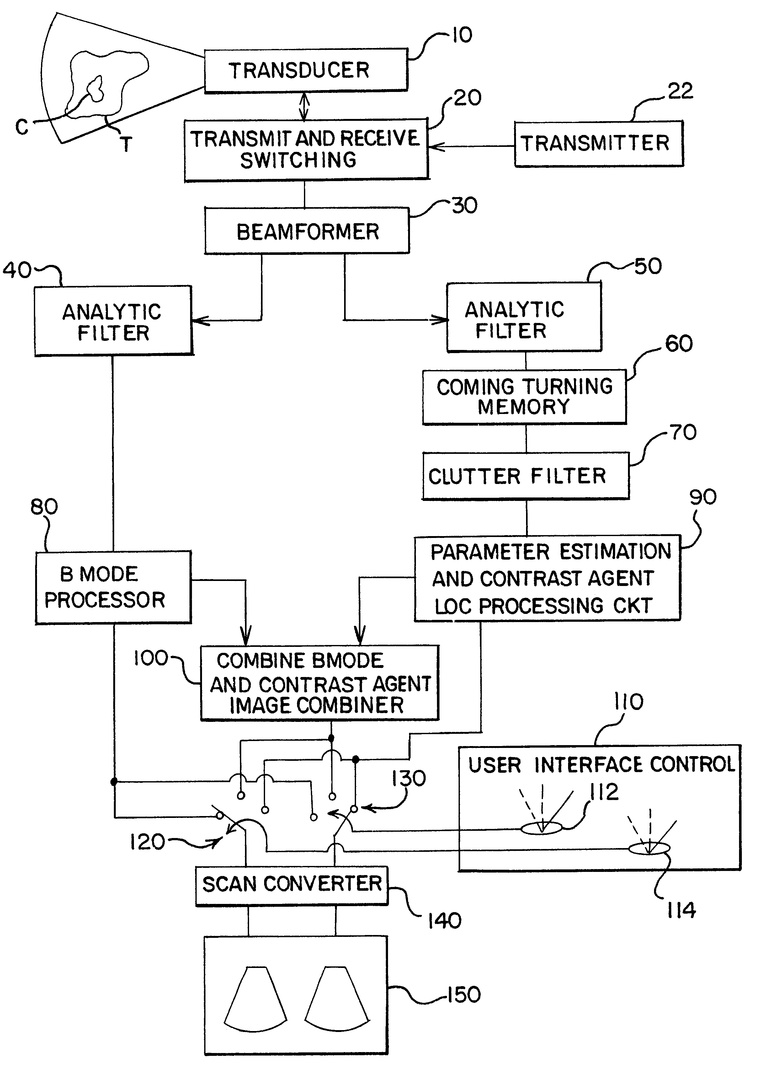
Turning now to the drawings, FIG. 1 shows a block diagram of a medical ultrasonic imaging system that incorporates a preferred embodiment of this invention. This imaging system includes a phased array ultrasonic transducer 10 that is coupled with transmit and receive switching circuitry 20. During transmit, the switching circuitry 20 connects a transmitter 22 to individual elements of the transducer array 10. The transmitter 22 applies excitation signals to the respective transducer elements to cause the transducer 10 to transmit and beam of ultrasonic energy into a tissue T comprising a contrast agent C. The contrast agent C can be any contrast agent that is suitable for use in the particular application. The contrast agent sold under the trade name Levovist can be taken as one example.

The tissue T and the contrast agent C generate echoes in response to each transmit pulse, and the transducer 10 generates echo signals that are connected by the switching circ...


PUM


Abstract

A medical ultrasonic imaging system transmits a set of two or more substantially identical transmit pulses into a tissue containing a contrast agent. The associated received pulses are filtered with a broadband filter that passes both the fundamental and at least one harmonic component of the echoes. The filtered received pulses are then applied to a clutter filter that suppresses harmonic and fundamental responses from slowly moving and stationary tissue, while clearly showing contrast agent response due to the loss of correlation effect. The disclosed system includes other signal paths for generating conventional B-mode images as well as combined images that include both components from the contrast-specific image as well as components from the B-mode image. An improved user interface allows the user to switch among these three images. Preferably the transmitter generates transmitted pulses having two or more spatially distinct focus zones, thereby improving the uniformity of contrast agent imaging over the imaged region.

Description

This invention relates to medical ultrasonic imaging, and in particular to systems for improved imaging of contrast agent included in imaged tissue.The nonlinear response of contrast agents such as microbubbles improves the detectability of microbubbles in tissue. Contrast agents effectively generate unique ultrasound signals such as second harmonic, third harmonic, subharmonic, or other harmonics that are not transmitted in significant quantities by a transducer. Good second harmonic contrast agents, in particular, have been shown to generate strong signals with minimal destruction of a population of contrast microbubbles.Another unique property of contrast agents is that they are disrupted by impinging sound waves. Ultrasound pulses in particular can move, crack, rearrange, split, and destroy microbubbles and their gas-encapsulating shells. When this type of disruption occurs, at least two received pulses will be decorrelated when at least two identical pulses are transmitted duri...

Claims


Application Information

Patent Timeline
Patent Type & Authority Patents(United States)
IPC IPC(8): A61B8/00G01S15/89G01S15/00G01S7/52A61B8/14
CPCA61B8/481G01S7/52041G01S7/52046G01S7/5206G01S7/52074G01S15/8981A61B8/14G01S7/52034
Inventor PHILLIPS, PATRICK J.GURACAR, ISMAYIL M.
Owner SIEMENS MEDICAL SOLUTIONS USA INC
Why Eureka
Try Eureka