
Color-coded medical tubes and post-insertion monitoring thereof

a color-coded, medical tube technology, applied in the direction of catheters, medical devices, other medical devices, etc., can solve the problems of post-insertion monitoring, serious medical complications, and additional discomfort of patients, and achieve the effect of reducing the problem of misplacemen

Inactive Publication Date: 2006-03-09
BETH ISRAEL DEACONESS MEDICAL CENT INC
View PDF14 Cites 16 Cited by
  • Summary
  • Abstract
  • Description
  • Claims
  • Application Information

AI Technical Summary

Benefits of technology

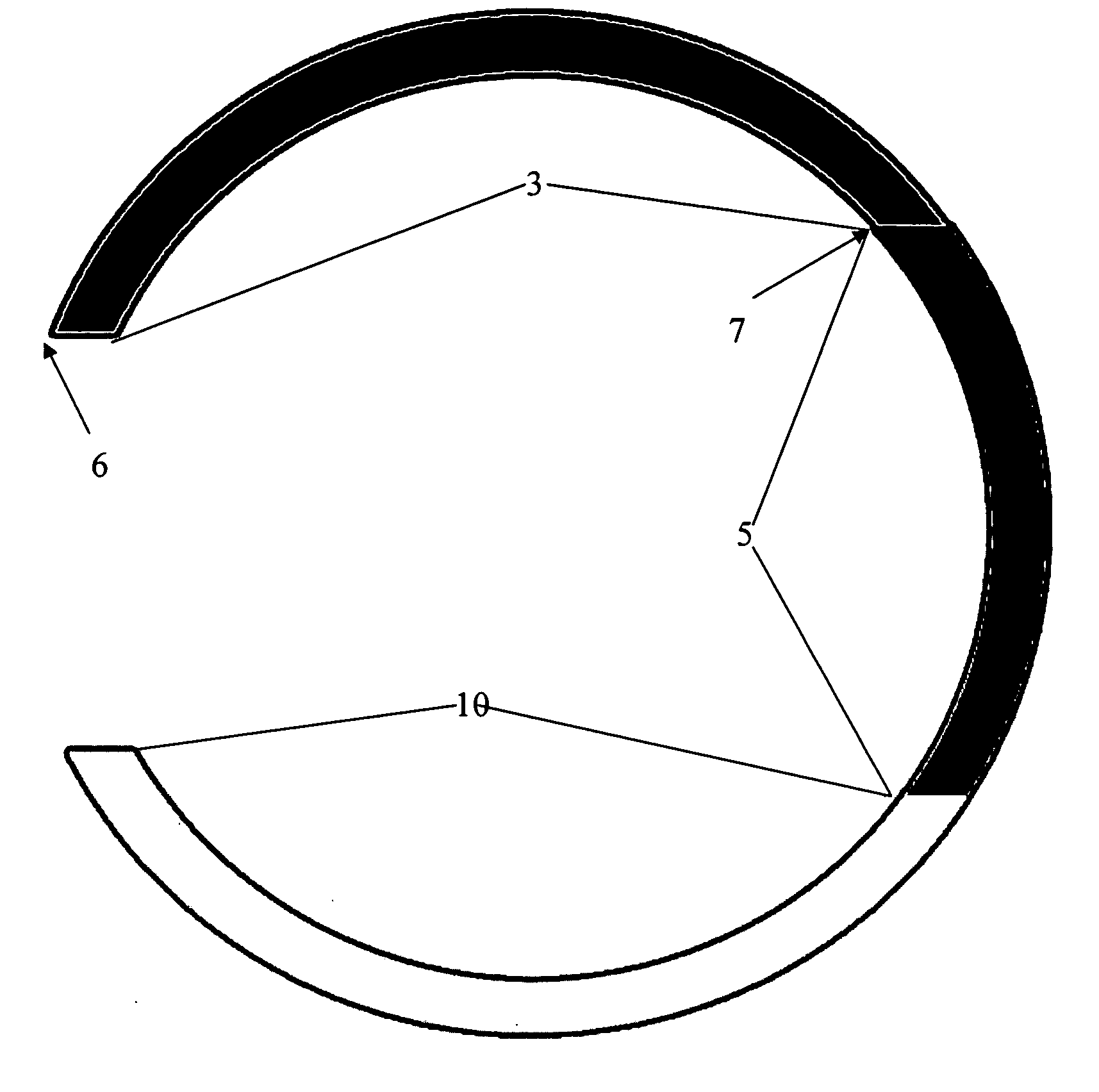
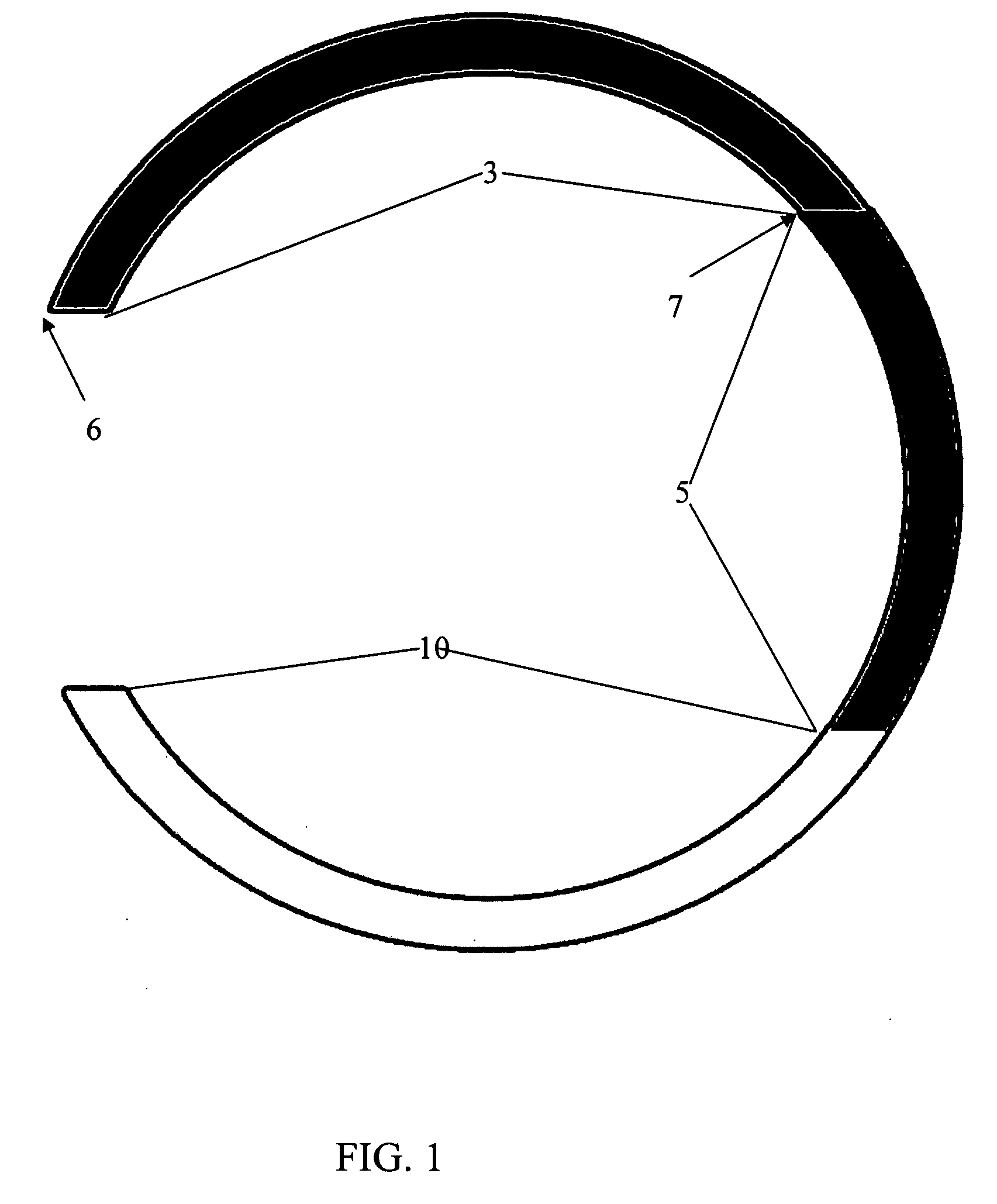
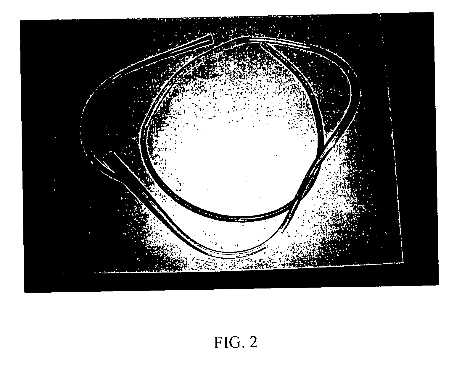
[0005] In a particular embodiment of the present invention, the medical tube comprises a tube body (made of the typical polymer / plastic as conventional medical tubes) with at least one color-coded warning zone section and at least one color-coded danger zone section. The warning zone section and the danger zone section are typically different, distinctive, easily identified colors. The tubes of the present invention can have multiple warning zone sections, and even danger zone sections, but typically have one warning zone and one danger zone. Insertion of the medical tube of the present invention is facilitated by visual identification of the color-coded sections as a guide to the insertion location within the patient. For example, with a nasogastric (NG) tube, about first 40 cm of the distal end of the tube is the danger zone section and is color-coded with a color such as red. The medical professional inserting the NG tube knows that the tube must be inserted into the patient at least as far as the color-coded danger zone section.
[0006] Color-coded sections such as a warning zone section and a danger zone section are also greatly advantageous for monitoring post-insertion problems with medical tubes. For example, if the warning zone section becomes visible post-insertion, even an untrained caregiver is alerted that the medical tube may need to be adjusted or reinserted. If the danger zone section becomes visible post-insertion, the caregiver or patient is alerted that the medical tube is in immediate need of adjustment or reinsertion and can contact their physician before serious medical damage can occur.
[0010] In yet another embodiment, the medical tube is a nasogastric tube comprising at least one visually distinguishable danger zone section wherein the danger zone section extends from the distal insertion tip to about 40 cm from the distal insertion tip along the tube body. The medical tube also has at least one visually distinguishable warning zone section wherein the warning zone section extends from the end of the danger zone section (at about 40 cm) to about 55 cm from the distal insertion tip along the tube body. It is appreciated by those of skill in the art that the lengths of such color-coded sections along a medical tube of the present invention will vary according to the specific use of the medical tube and that the lengths of color-coded divisions for different specific purposes are easily determined. It is also appreciated by those of skill in the art that the color-coded zones of the medical tubes for the pediatric population will be proportionally smaller.
[0013] There are several advantages to the medical tubes of the present invention. First, the tube is not functionally altered in any way. The color-coded medical tube does not require external devices such as magnets or radio transmitters in order to monitor insertion or placement. Second, professionals inserting the tube are aided by the color-coding to determine the position of the tube in the patient's body. This will reduce problems of misplacement by even highly trained medical professionals. Third, once the tube is functioning, any change in the tube's position / location in the patient will be clearly visible thus indicating that correction may be, or is, required.

Problems solved by technology

Most medical professionals who insert such tubes are intensely trained and highly skilled in these techniques, but nonetheless, there are problems.
Inappropriate insertion can lead to serious medical complications, even death.
Often proper insertion of medical tubes must be confirmed or monitored by x-ray causing additional discomfort to the patient.
Even if a medical tube is initially inserted properly, one of the biggest challenges for a medical professional is post-insertion monitoring.
Often, medical tubes that are inserted properly are deliberately pulled at by an uncomfortable patient, or dislocated by accident.
The problem of post-insertion monitoring is even more apparent with the increase in home care procedures for patients.
In such situations a medical professional is not at home with the patient and the burden of monitoring proper tube insertion falls on the patient or an untrained caregiver.
Currently there is no way to easily recognize when a medical tube becomes misplaced after the initial insertion.

Method used


Image

  • Color-coded medical tubes and post-insertion monitoring thereof
  • Color-coded medical tubes and post-insertion monitoring thereof
  • Color-coded medical tubes and post-insertion monitoring thereof

Examples


Embodiment Construction

[0018] A description of preferred embodiments of the invention follows.

[0019] The present invention pertains to a medical tube, a method of inserting the medical tube and a method for post-insertion monitoring of the medical tube. As used herein the term “medical tube” means any type of tube which may be inserted into a patient's body and is expected to stay undisturbed within the patient, including, but not limited to, nasogastric tubes, chest tubes, feeding tubes, drains, foley catheters and central lines.

[0020] The medical tube of the present invention has color-coded sections. The terms “color-coded” and “color” as used herein mean any visual way of distinguishing different sections along the tube body. This includes differentiation based on color and / or luminescence such as phosphorescence or fluorescence. For example, sections of the tube can be of distinct colors such as red or green. Sections can also be luminescent sections, either alone instead of a color, or in addition...


PUM


Abstract

The present invention describes color-coded medical tubes and methods of monitoring the insertion and post-insertion of the same.

Description

BACKGROUND OF THE INVENTION [0001] During the course of an illness patients often require the insertion of medical tubes for a variety of purposes. Such medical tubes include, nasogastric tubes, chest tubes, feeding tubes, drains, foley catheters and central lines. For example, a nasogastric (NG) tube inserted through the nose, down the esophagus and into the stomach can be used for removing the stomach fluids. Clearly proper insertion and monitoring the placement / location of these tubes in the patient is critical. Most medical professionals who insert such tubes are intensely trained and highly skilled in these techniques, but nonetheless, there are problems. Inappropriate insertion can lead to serious medical complications, even death. [0002] Presently, medical tubes that are inserted into patients are made of either clear / colorless polymers, or are of a single color. Some medical tubes have intervals marked with a thin black line, but these lines are not clearly noticeable and, o...

Claims


Application Information

Patent Timeline
IPC IPC(8): A61M25/00
CPCA61M25/00A61M39/08A61M2205/6081A61M2205/583A61M2025/0008
Inventor PATEL, ASHISH R.
Owner BETH ISRAEL DEACONESS MEDICAL CENT INC
Learn More