
Registration of scanning data acquired from different patient positions

a scanning data and patient position technology, applied in the field of volume based three-dimensional virtual examination, can solve the problems of operator not viewing, incomplete control of a camera in a large domain, and not giving the viewer the flexibility to alter the course or investiga

Inactive Publication Date: 2006-03-23
THE RES FOUND OF STATE UNIV OF NEW YORK
View PDF2 Cites 16 Cited by
  • Summary
  • Abstract
  • Description
  • Claims
  • Application Information

AI Technical Summary

Benefits of technology

The present patent describes a method and system for generating a three-dimensional visualization of an object, such as a human organ, for examination and treatment purposes. The method involves acquiring image data of the object and converting it into a volume data set. The volume data set is then used to create a fly-path through the object, allowing the operator to examine it from different angles and positions. The fly-path can be generated based on volume shrinking from the wall of the object or by partitioning the object into segments and connecting the points. The system includes an imaging scanner, processor, and display for the virtual examination. The method and system can be used for identifying abnormal features in the object and facilitating the diagnosis and treatment of colon polyps and other abnormalities.

Problems solved by technology

However, complete control of a camera in a large domain would be tedious and tiring, and an operator might not view all the important features between the start and finishing point of the exploration.
However, this second technique does not give the viewer the flexibility to alter the course or investigate an interesting area viewed along the flight path.
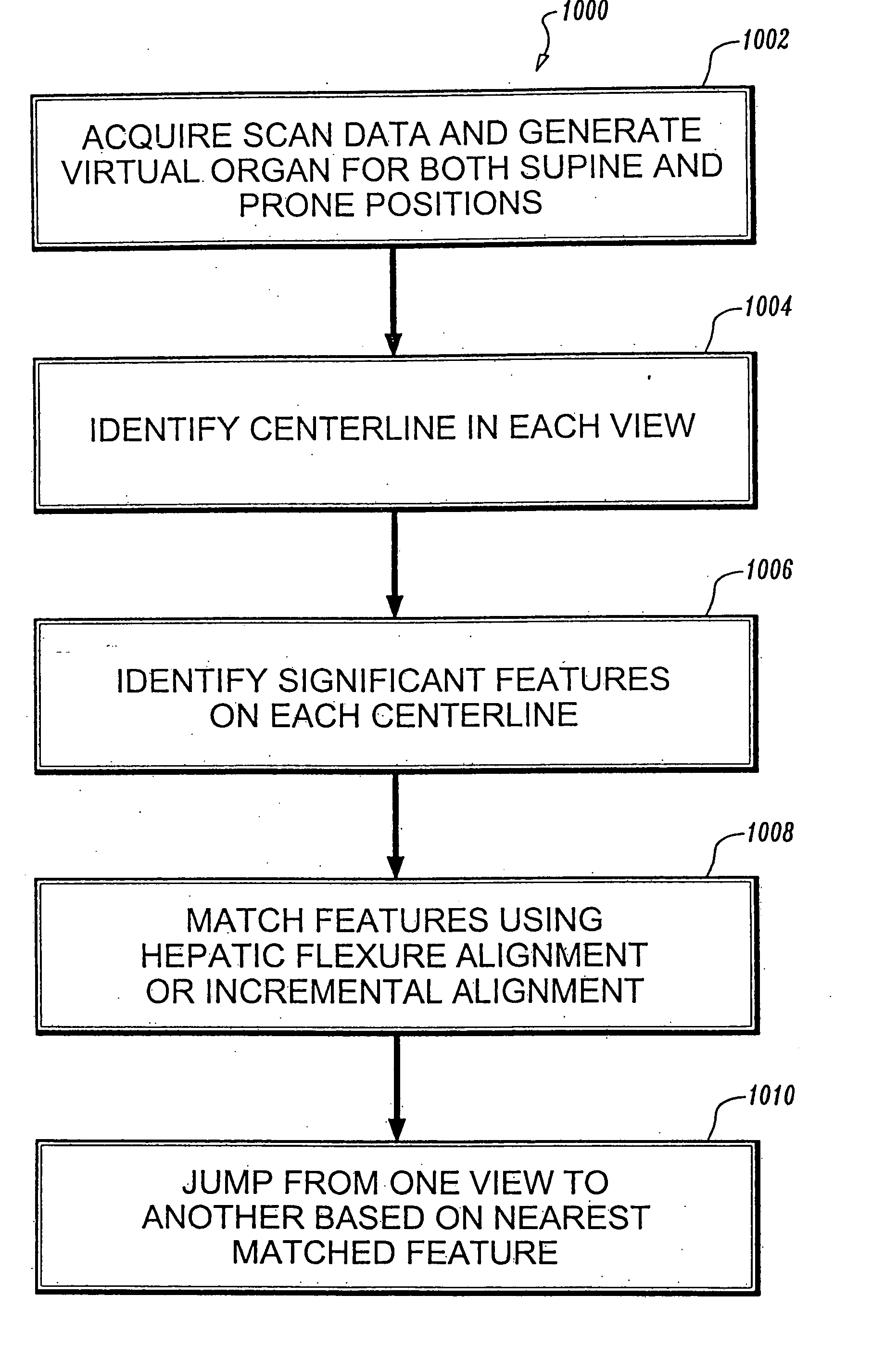
As a consequence, when an interesting feature is located in one orientation or view, there is currently no way to automatically “jump” to that same feature in the other orientation or view.

Method used


Image

  • Registration of scanning data acquired from different patient positions
  • Registration of scanning data acquired from different patient positions
  • Registration of scanning data acquired from different patient positions

Examples


Embodiment Construction

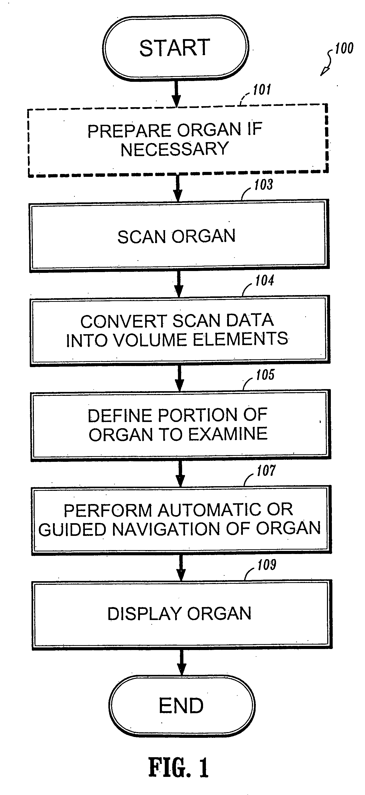
[0032] While the methods and systems described herein may be applied to any object to be examined, the preferred embodiment to be described is the examination of an organ in the human body, specifically the colon. The colon is long and twisted, which makes it especially suited for a virtual examination saving the patient both monetary expense as well as the discomfort and increased hazard of a physical probe. Other examples of organs that can be examined include the lungs, stomach and portions of the gastro-intestinal system, the heart and blood vessels.

[0033] As shown in FIG. 1, a method for performing a virtual examination of an object such as a colon is indicated generally by the reference numeral 100. The method 100 illustrates the steps necessary to perform a virtual colonoscopy using volume visualization techniques. Step 101 prepares the colon to be scanned in order to be viewed for examination if required by either the doctor or the particular scanning instrument. This prepa...


PUM


Abstract

A system and method are provided for generating a three-dimensional visualization image of an object such as an organ using volume visualization techniques and exploring the image using a guided navigation system that allows the operator to travel along a flight path and to adjust the view to a particular portion of the image of interest in order, for example, to identify polyps, cysts or other abnormal features in the visualized organ; where imaging data sets are acquired using conventional two-dimensional scans of the object in both a supine and prone orientation, and correspondence between the respective data sets is determined to permit jumping from one visualization orientation to the other while remaining at the same virtual location within the organ.

Description

CROSS-REFERENCE TO RELATED APPLICATION [0001] This application is a continuation of application Ser. No. 10 / 496,430 filed on May 21, 2004, which claims the benefit under 35 U.S.C. § 365(c) of PCT International Application Designating the United States, Serial No. PCT / US02 / 37387, entitled “REGISTRATION OF SCANNING DATA ACQUIRED FROM DIFFERENT PATIENT POSITIONS”, and filed Nov. 21, 2002, which is incorporated herein by reference in its entirety; and which, in turn, claims the benefit under 35 U.S.C. § 119(e) of U.S. Provisional Patent Application Ser. No. 60 / 331,712, entitled “New Features For Virtual Colonoscopy”, filed Nov. 21, 2001, which is incorporated herein by reference in its entirety.BACKGROUND OF THE INVENTION [0002] The present disclosure relates to a system and method for performing a volume based three-dimensional virtual examination, and more particularly relates to a method providing enhanced visualization and navigation properties. [0003] Two-dimensional (“2D”) visuali...

Claims


Application Information

Patent Timeline
Patent Type & Authority Applications(United States)
IPC IPC(8): G06K9/00G06T7/00G06T15/08G06T17/00
CPCG06T7/0012G06T7/0028G06T7/0038G06T7/60G06T15/08G06T2207/30028G06T2207/10081G06T2207/10088G06T2207/20044G06T2207/30004G06T2200/04Y10S128/92G06T7/33G06T7/38
Inventor YOAKUM-STOVER, SUZANNEKAUFMAN, ARIE E.KREEGER, KEVIN
Owner THE RES FOUND OF STATE UNIV OF NEW YORK
Learn More