Apparatus for non-invasive determination of direction and rate of change of an analyte

a fluid analyte and non-invasive technology, applied in the field of non-invasive methods and apparatus for measuring fluid analyte, can solve the problems of inability to monitor glucose at home, repeated irritation and inconvenience, and discourage vigilant self-monitoring, so as to improve the stability and accuracy of robinson measurement, and improve the accuracy of non-invasive measurement. , the effect of increasing blood flow

Inactive Publication Date: 2006-07-27
INLIGHT SOLUTIONS
View PDF7 Cites 27 Cited by
  • Summary
  • Abstract
  • Description
  • Claims
  • Application Information

AI Technical Summary

Benefits of technology

[0023] The present invention includes methods and apparatuses for non-invasively measuring several characteristics of analyte concentrations in tissue. Some embodiments enable measurement of the value, rate of change, and direction of change of the analyte concentration by analyzing tissue. Several embodiments use spectroscopic techniques to achieve the non-invasive measurements, and can incorporate means for accelerating the equilibration of the concentrations of specific analytes between tissue compartments in a sample area to provide a present analysis or to amplifying the ability to monitor changes.
[0026] The first means for transmitting light, in several embodiments, includes placing the energy source adjacent to the input element so that light from the source propagates through the air to the input element. The second means for transmitting light can include a single mirror or system of mirrors that direct the light energy exiting the output element through the air to the spectrum analyzer. Alternative embodiments can include using a transmission medium, such as glass, plastics or other materials, instead of air to prevent reflections or absorption in the air, or simply to provide a sealed, more robust system.
[0031] Before the output element can receive the light emanating from the tissue, the light exits the tissue and enter the output element. Again, there can be reflections at the interfaces of the tissue and any medium between the tissue and the output element. These reflections, along with diffraction and absorption by any medium between the tissue and the output element, can reduce the intensity received by the output element and, where light is not incident normally on one of the interfaces, can create problems of internal reflection.
[0033] In some embodiments of the present invention, it can be useful to accelerate the equilibration of the analyte concentrations in these separate tissue compartments. Such acceleration can improve the ability of the sensor element to detect, without requiring inordinately high sensitivity, a rate of change of the analyte concentration in the vascular system. In preferred embodiments, this acceleration toward equilibration is effected by causing an increase in vascular flow, for example, by applying localized heating of the tissue.
[0036] Analysis of the received data can also be used to predict the direction and rate at which blood glucose concentrations are changing. In some embodiments, multiple readings can be captured by the sensor element and compared to one another in order to determine the direction and rate of change of blood glucose concentrations. The speed at which change is taking place, in some embodiments, is used to predict a future value of blood glucose concentration. In other embodiments, the speed at which the change is taking place is used to predict the present concentration of the analyte in other compartments, such as blood. In several embodiments, the measurement of direction and rate of change of glucose concentration is enhanced by the use of an equilibration accelerating means, for example, localized heating, an applied rubrifractant, or an ingested or injected vasodilator.

Problems solved by technology

Presently, most home glucose monitoring is limited to methods and systems requiring a lancet cut into the finger.
Repeated lancet cuts lead to repeated irritation and inconvenience, discouraging vigilant self-monitoring.
This is believed so compromising to the diabetic patient that the most effective use of any form of diabetic management is rarely achieved.
However, non-invasive analyte detection in blood in this region is problematic, if not impossible, because water makes up the majority of the blood and demonstrates strong absorption characteristics at mid-infrared wavelengths.
Due to the overlapping nature of these absorption bands, reliable non-invasive glucose measurement would be very difficult if only a single frequency were used for analysis.
An additional difficulty can arise with respect to the incident power required to achieve a readable signal with respect to glucose levels.
One way to achieve higher sensitivity is to use a relatively large input power, which, however, can cause non-linearities in the optical systems themselves as well as in the material subjected to optical radiation.
However, a problem encountered in non-invasive skin based measurements of standard medical blood analytes is the disparity between the concentration of a given analyte in the blood and the same analyte in the adjacent skin tissue water or interstitial fluid.
Thus, the fact that the glucose must move between the tissue and blood causes errors in both interstitial space glucose and total tissue glucose concentration measurements when used as surrogates for blood glucose concentration measurements.
This presents obvious problems with respect to using the surrogate methods for monitoring and guiding therapy in diabetic patients.
Given the concentration difference, determining whether a given technique is working based on infrequent, discrete measurements is believed impossible.
Without more frequent measurements, it is difficult to determine whether the patient's blood glucose is in a steady state condition or is in flux.
If a tissue measurement were made it would inappropriately report a level that is higher than the actual blood glucose concentration and that would not inform the patient of the quickly changing concentration.
The result of very low blood glucose concentrations (below 40 mg / dl, 2.2 mmol) is often loss of consciousness, coma and even brain damage or death if the patient is not discovered in time for medical intervention.
An additional problem exists with respect to measuring changing blood glucose levels.
However, just like the first blood draw, the second blood draw would be another discomforting inconvenience; the additional discomfort and inconvenience can further discourage patient self-monitoring.
Similar problems exist with regards to measurement of other analytes.
Current measurement methods cannot detect these circumstances.

Method used


Image

  • Apparatus for non-invasive determination of direction and rate of change of an analyte
  • Apparatus for non-invasive determination of direction and rate of change of an analyte
  • Apparatus for non-invasive determination of direction and rate of change of an analyte

Examples


Embodiment Construction

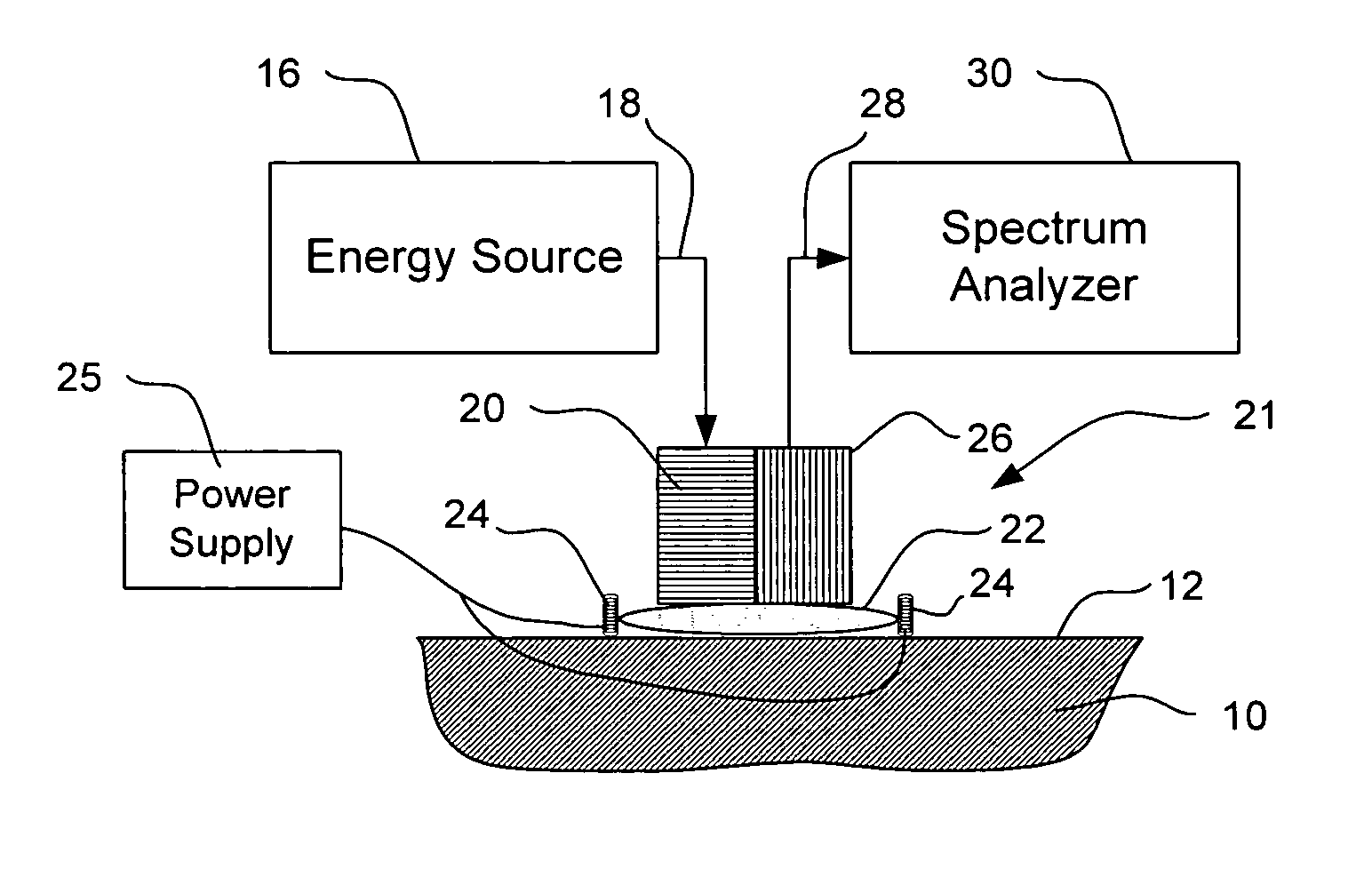
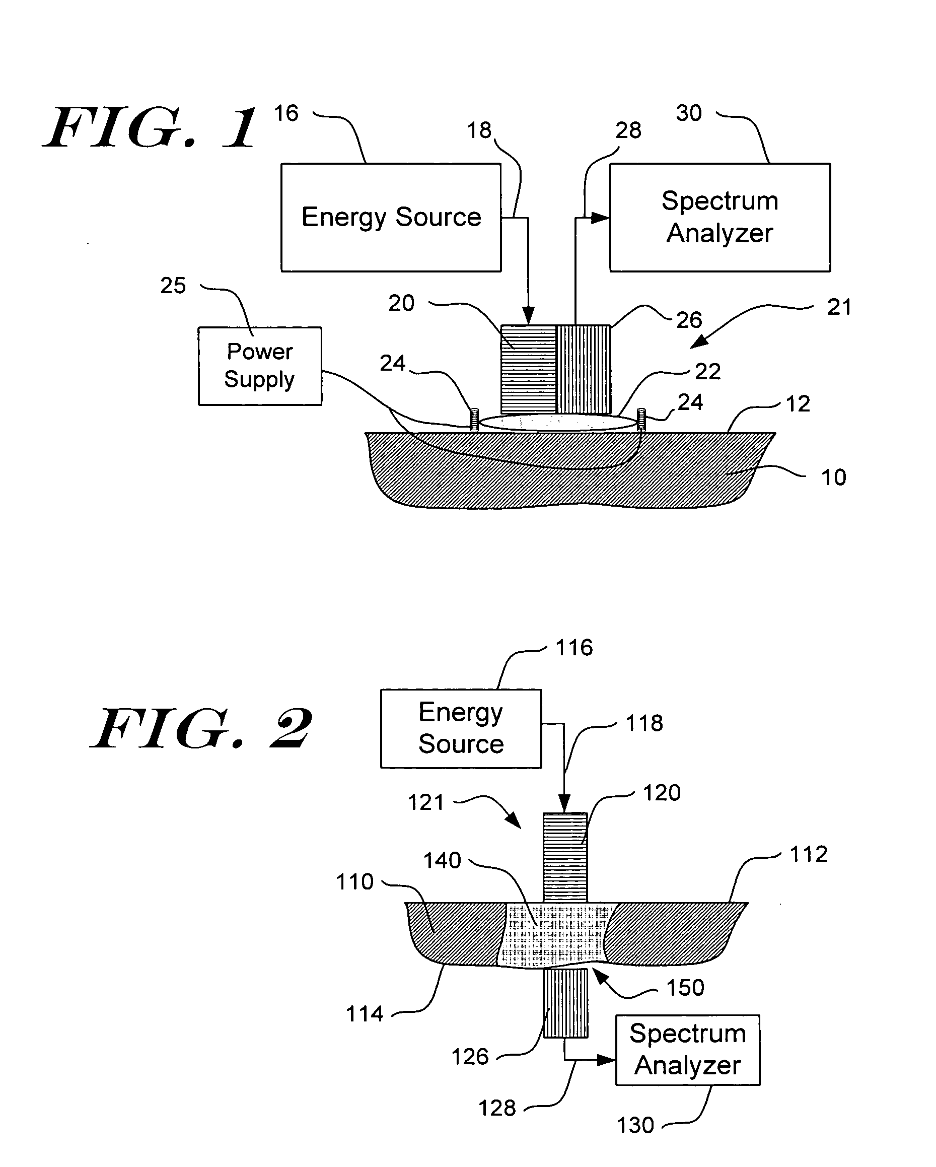
[0045] Detailed embodiments of the present invention are disclosed herein. However, it is to be understood that the disclosed embodiments are merely exemplary of the present invention which can be embodied in various systems. Therefore, specific details disclosed herein are not to be interpreted as limiting, but rather as a basis for the claims and as a representative basis for teaching one of skill in the art to variously practice the invention. The invention makes use of signals, described in some of the examples as absorbance or other spectroscopic measurements. Signals can comprise any measurement obtained concerning the tissue, e.g., absorbance, reflectance, intensity of light returned, fluorescence, transmission, Raman spectra, all at one or more wavelengths; concentration of an analyte, and characteristics relating to change of any of the preceding. The invention also concerns analyte properties, where an analyte property can include, as examples, analyte concentration, prese...


PUM

PropertyMeasurementUnit
wavelengthsaaaaaaaaaa
concentrationsaaaaaaaaaa
temperatureaaaaaaaaaa

Abstract

The present invention relates generally to a non-invasive method and apparatus for measuring a fluid analyte, particularly relating to glucose or alcohol contained in blood or tissue, utilizing spectroscopic methods. More particularly, the method and apparatus incorporate means for detecting and quantifying changes in the concentration of specific analytes in tissue fluid. Also, the method and apparatus can be used to predict future levels of analyte concentration either in the tissue fluid or in blood in an adjacent vascular system.

Description

CROSS REFERENCES TO RELATED APPLICATIONS [0001] This application is a continuation of and claims priority under 35 U.S.C. § 120 to U.S. patent application Ser. No. 10 / 753,506, entitled “Non-Invasive Determination of Direction And Rate Of Change of an Analyte,” filed Jan. 8, 2004; which was a continuation-in-part of U.S. patent application Ser. No. 09 / 864,774, entitled “Method and Apparatus for Non-Invasive Blood Analyte Measurement with Fluid Compartment Equilibration,” filed May 24, 2001, which was a continuation of U.S. Pat. No. 6,240,306, which was a Continuation-in-Part of U.S. patent application Ser. No. 09 / 174,812, filed Oct. 19, 1998 entitled “Method for Non-Invasive Blood Analyte Measurement with Improved Optical Interface”, which is a Continuation-in-Part of U.S. patent application Ser. No. 08 / 844,501, filed Apr. 18, 1997, entitled “Method for Non-Invasive Blood Analyte Measurement with Improved Optical Interface”, now U.S. Pat. No. 5,823,951, issued Oct. 20, 1998, which is...

Claims


Application Information

Patent Timeline
Patent Type & Authority Applications(United States)
IPC IPC(8): A61B5/00G01N
CPCA61B5/0059A61B5/0075A61B5/14532A61B5/1455A61B5/1491A61B2560/0233A61B2562/146
Inventor GARDNER, CRAIG M.RIDDER, TRENT D.GRUNER, WILLIAM
Owner INLIGHT SOLUTIONS
Why Eureka
Try Eureka