Method of database-guided segmentation of anatomical structures having complex appearances

a technology of anatomical structure and database, applied in image analysis, image enhancement, medical science, etc., can solve the problems of inability to capture complex structure appearance, limited applicability of underlying assumptions, and difficulty in automated segmentation of echocardiographic images

Active Publication Date: 2006-08-03
SIEMENS HEALTHCARE GMBH
View PDF25 Cites 67 Cited by
  • Summary
  • Abstract
  • Description
  • Claims
  • Application Information

AI Technical Summary

Problems solved by technology

These approaches are limited by the validity of the underlying assumptions and cannot capture complex structure appearance.
Automated segmentation of echocardiographic images has proved to be challenging due to large amounts of noise, signal drop-out and large variations between appearance, configuration and shape of the left ventricle.
However, as the complexity of targeted segmentation increases, it is more difficult to encode prior knowledge into the grouping task.

Method used


Image

  • Method of database-guided segmentation of anatomical structures having complex appearances
  • Method of database-guided segmentation of anatomical structures having complex appearances
  • Method of database-guided segmentation of anatomical structures having complex appearances

Examples


Embodiment Construction

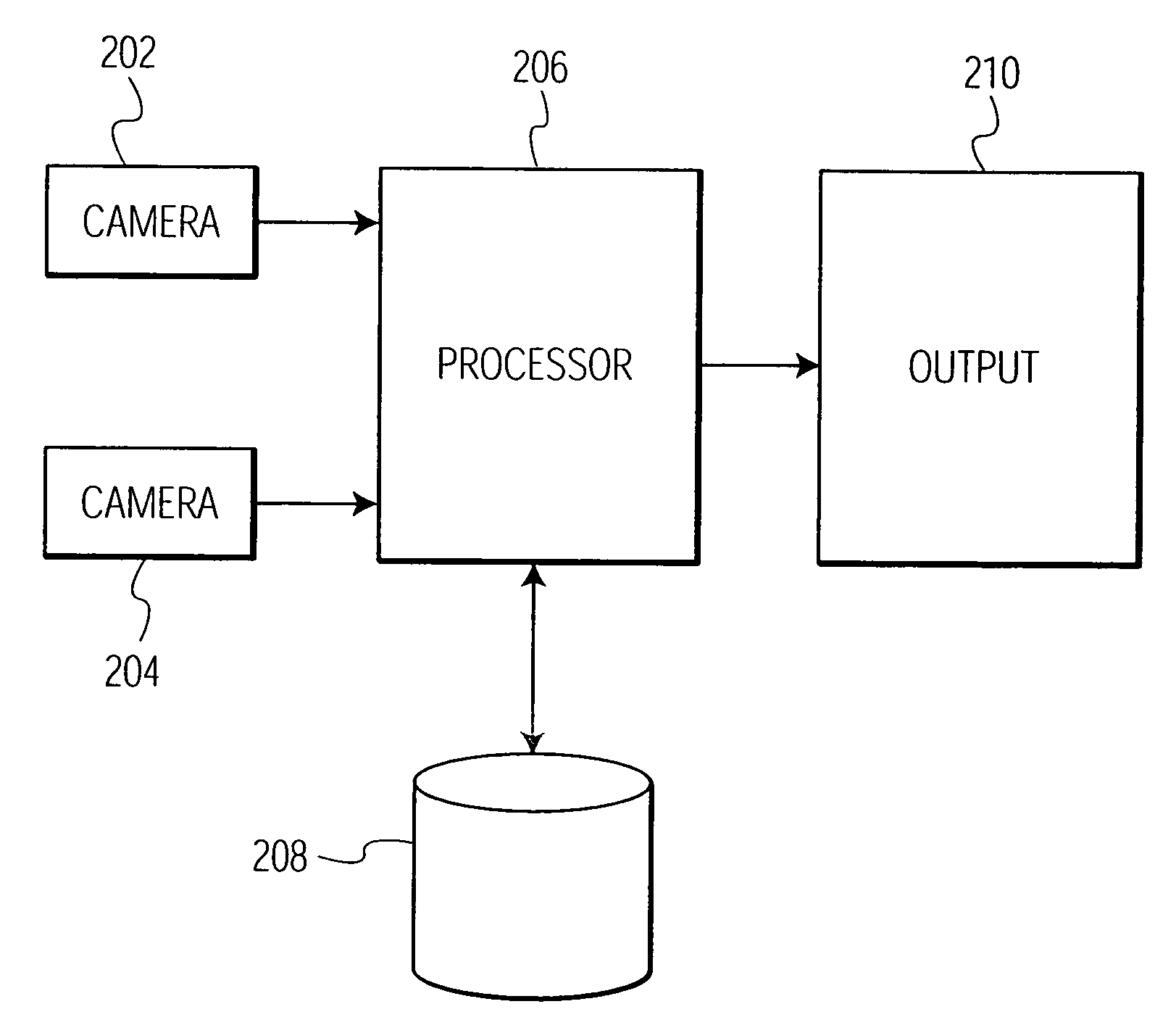
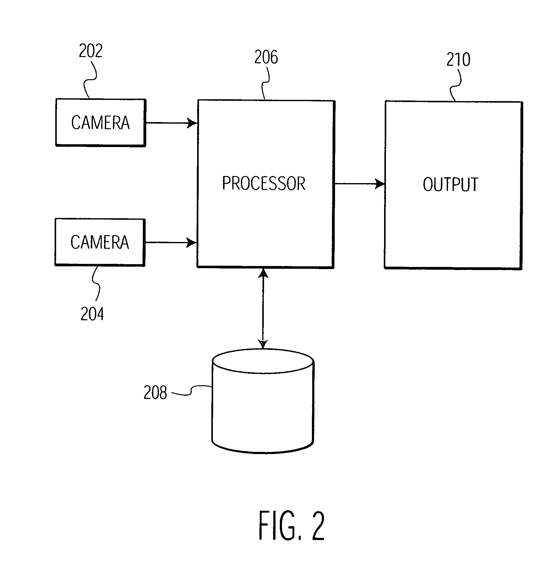
[0028] The present invention is directed to a method for detecting and matching anatomical structures. An example where such a method would be utilized is for to detecting regional wall motion abnormalities in the heart by detection and segmentation of the ventricle endocardial or epicardial borders through machine learning, or classification, and by identifying similar cases from annotated databases. It is to be understood by those skilled in the art that the present invention may be used in other applications where shape detection and matching is useful such as, but not limited to, recognizing human features such as facial features or other body features. The present invention can also be used in 2 dimensional, 3 dimensional and 4 dimensional (3D+time) data analysis, such as medical analysis of anatomical structures such as the heart, lungs or tumors, which can be evolving over time.

[0029] For purposes of describing the present invention, an example will be described for database...


PUM


Abstract

A method for segmenting an anatomical structure of interest within an image is disclosed. The anatomical structure of interest is compared to a database of images of like anatomical structures. Those database images of like anatomical structures that are similar to the anatomical structure of interest are identified. The identified database images are used to detect the anatomical structure of interest in the image. The identified database images are also used to determine the shape of the anatomical structure of interest. The anatomical structure of interest is segmented from the image.

Description

CROSS-REFERENCE TO RELATED APPLICATION [0001] This application claims the benefit of U.S. Provisional Patent Application Ser. No. 60 / 625,908, filed on Nov. 8, 2004, which is incorporated by reference in its entirety.FIELD OF THE INVENTION [0002] The present invention is directed to a method for detecting structure and shape inference of an anatomical structure using database-guided segmentation, and more particularly, to a method for detecting structure and shape inference of an anatomical structure by first discriminating the structure from the background and using discriminative features to infer the shape of the structure. BACKGROUND OF THE INVENTION [0003] Segmentation of anatomical structures has been traditionally formulated as a perceptual grouping task and solved through clustering and variational approaches. However, such strategies require a priori knowledge to be explicitly defined in the optimization criterion (e.g., “high-gradient border”, “smoothness” or “similar inten...

Claims


Application Information

Patent Timeline
Patent Type & Authority Applications(United States)
IPC IPC(8): G06K9/34G06K9/00G06V10/772
CPCG06K9/6255G06T7/0081G06T2207/10132G06T2207/20081G06T2207/30048Y10S128/922Y10S128/923G06T7/11G06V10/772G06F18/28
Inventor GEORGESCU, BOGDANZHOU, XIANGCOMANICIU, DORINRAO, R. BHARATGUPTA, ALOK
Owner SIEMENS HEALTHCARE GMBH
Why Eureka
Try Eureka