Method for expanding the domain of imaging software in a diagnostic work-up

a diagnostic work-up and imaging software technology, applied in the field of medical imaging data analysis, can solve the problems of not providing an accurate way to detect secondary conditions, medical images, and no system compensates for differences in accuracy

Inactive Publication Date: 2006-10-26
YANKELEVITZ DAVID DR +1
View PDF29 Cites 25 Cited by
  • Summary
  • Abstract
  • Description
  • Claims
  • Application Information

AI Technical Summary

Problems solved by technology

Unfortunately, such known methods do not provide an accurate way to detect secondary conditions that may present in a medical image initially acquired for another condition, other than the condition that is the subject of t

Method used


Image

  • Method for expanding the domain of imaging software in a diagnostic work-up
  • Method for expanding the domain of imaging software in a diagnostic work-up
  • Method for expanding the domain of imaging software in a diagnostic work-up

Examples


Embodiment Construction

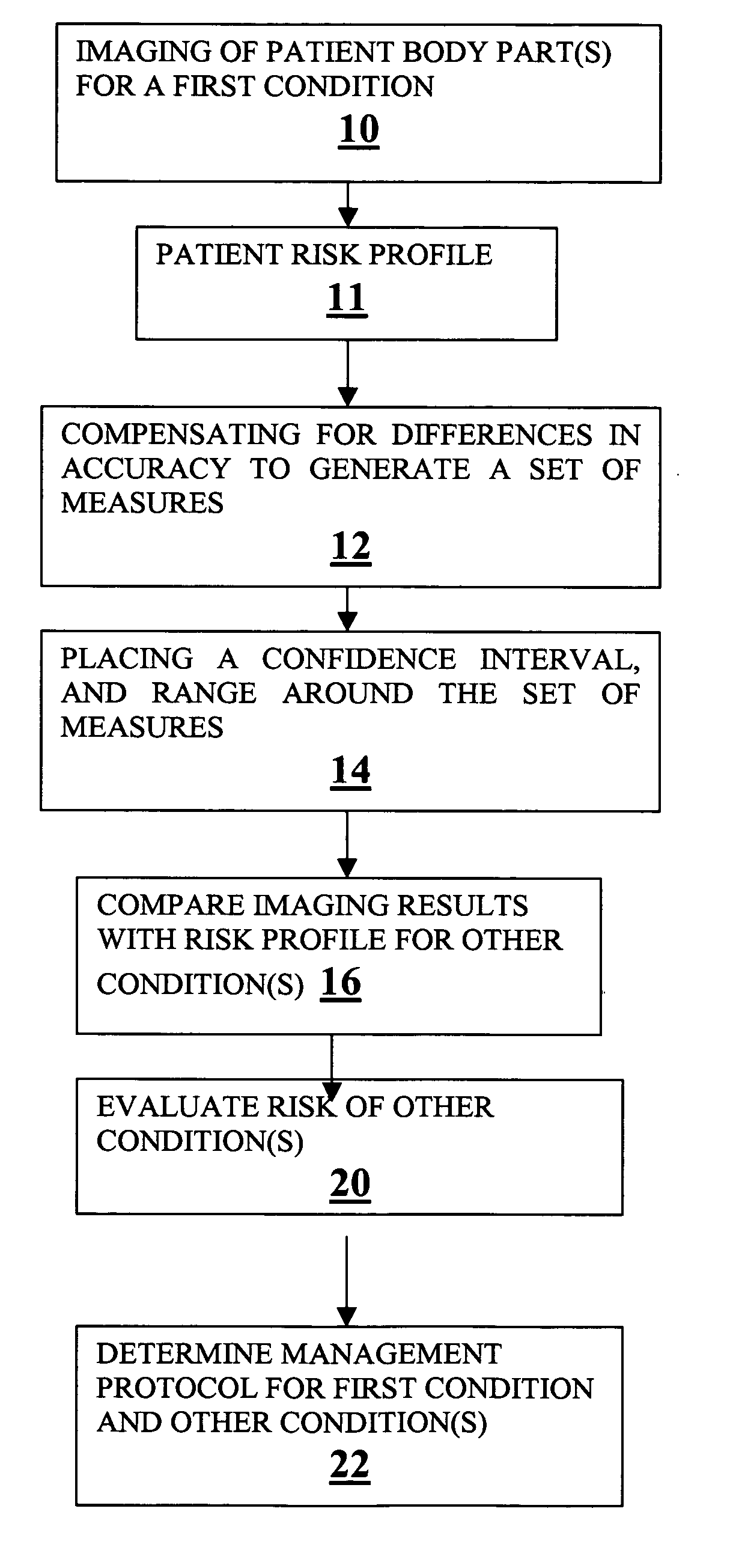
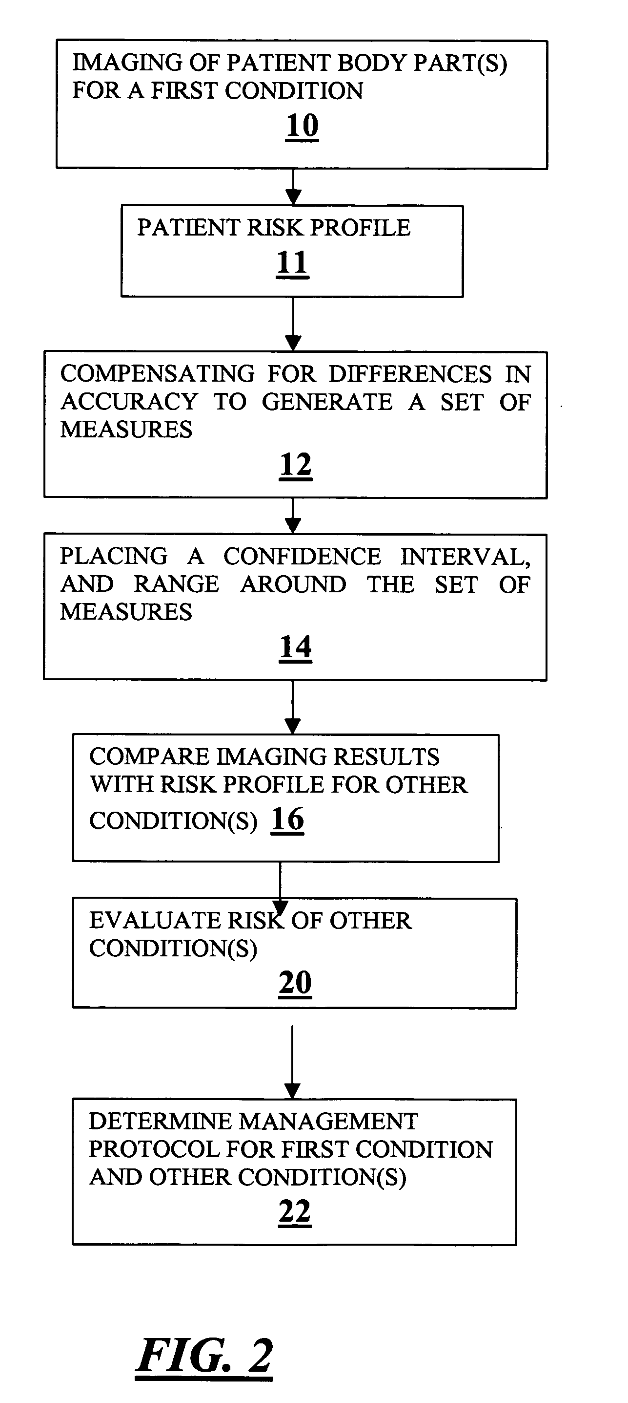
[0018] Preliminarily, it should be noted that while a particular system and method described in detail herein is for analyzing medical imaging data, such as radiology data, this is not by way of limitation, but solely for the purposes of illustration, and the invention may also be employed for analyzing data of other types. (the clinical information that is used to suggest the risk profile of the patient can come from a variety of sources, i.e. age, smoking history, blood profiles, genetic characterizations etc)

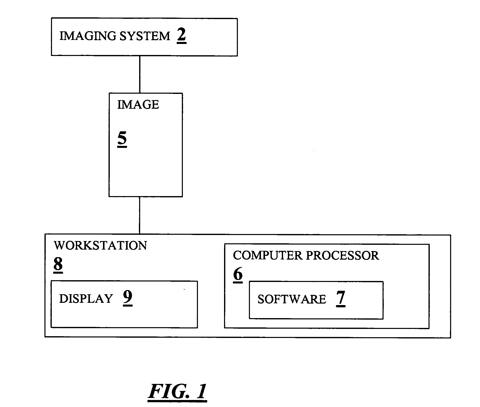
[0019] Referring now to FIG. 1, there shown is a simplified block diagram of a system for expanding the domain of imaging software in a diagnostic work-up constructed in accordance with one embodiment of the present invention. An imaging system 2 produces image data 5. The image data is processed in a workstation 8. The workstation 8 preferably includes a computer processor 6 running image processing software 7, and a display 9.

[0020] The medical imaging system 2 may advant...


PUM


Abstract

A method for expanding the domain of imaging software in a diagnostic work-up includes the steps of: imaging a patient's body parts for a first condition using a first imaging technique directed to a first condition; acquiring known patient risk factors indicating a second condition; compensating for differences in accuracy between a first imaging technique directed to a first condition, and a second imaging technique directed to testing for the second condition to generate a set of measures; placing a range and confidence interval around the set of measures; and evaluating for a second condition using the set of measures and known patient risk factors.

Description

CROSS-REFERENCE TO RELATED APPLICATION [0001] This application claims priority under 35 U.S.C. § 119(e) to U.S. Provisional Patent Application No. 60 / 662,190, filed Mar. 16, 2005, which is incorporated herein by reference.FIELD OF THE INVENTION [0002] The present invention relates generally to analysis of medical imaging data, and, more particularly to a method for expanding the domain of imaging software in a diagnostic work-up. BACKGROUND OF THE INVENTION [0003] Imaging protocols with modern CT scanners and other imaging equipment now allow for high resolution exam images to be obtained for various parts of the body within short time intervals. The resolution on these routine examinations is now similar in quality to the protocols used for screening or diagnostic protocols used to find specific conditions. In addition, computer-aided detection (CAD) software helps radiologists read medical images such as mammograms and computed tomography (also known as CT or CAT scanning) chest s...

Claims


Application Information

Patent Timeline
IPC IPC(8): G06K9/62A61B5/05G16H30/20G16H30/40G16H50/30G16H70/20
CPCA61B6/12G06F19/321G06T2207/30004G06T7/0012G06F19/345G16H50/20G16H50/30G16H70/20G16H30/20G16H30/40
Inventor YANKELEVITZ, DAVID F.REEVES, ANTHONY P.HENSCHKE, CLAUDIA INGRID
Owner YANKELEVITZ DAVID DR
Why Eureka
Try Eureka