
System and method for performing a three-dimensional virtual examination of objects, such as internal organs

a three-dimensional virtual examination and object technology, applied in the field of system and method for performing a volume-based three-dimensional virtual examination, can solve the problems of invasive, inconvenient, and difficult to store data, and achieve the effect of efficient storage and recall of data

Inactive Publication Date: 2007-05-10
THE RES FOUND OF STATE UNIV OF NEW YORK
View PDF58 Cites 101 Cited by
  • Summary
  • Abstract
  • Description
  • Claims
  • Application Information

AI Technical Summary

Benefits of technology

The invention is a method for generating a three-dimensional visualization image of an object using volume visualization techniques and exploring it using a guided navigation system. This allows the operator to identify abnormal features in the object by performing virtual examination. The method involves acquiring imaging scan data of the object and converting it to a plurality of voxels. Image segmentation is then performed to classify the voxels into different categories. An expanded dataset is generated by interpolating between the voxels and a reduced resolution dataset is generated for efficient storage and recall. The method can be used for performing virtual angiography to detect and monitor the progression of aneurysms and determine the characteristics needed to place a stent graft. Additionally, the method can be used to define a skeleton for a three-dimensional image representation of a hollow object.

Problems solved by technology

Colon cancer continues to be a major cause of death throughout the world.
The colonoscopy, although reliable, is both relatively costly in money and time, and is an invasive, uncomfortable painful procedure for the patient.
However, barium enemas may not always provide a view of the entire colon, require extensive pretreatment and patient manipulation, is often operator-dependent when performing the operation, exposes the patient to excessive radiation and can be less sensitive than a colonoscopy.
Another leading cause of cancer deaths in the United States is bladder cancer.
However, such tests are generally only effective at detecting bladder cancer in its later developed stages and does not provide any information regarding the size or location of a cancerous growth.
However, cystoscopy is an invasive procedure which offers a physician a limited field of view and lacks an objective indication of size.
However, complete control of a camera in a large domain would be tedious and tiring, and an operator might not view all the important features between the start and finishing point of the exploration.
However, this second technique does not give the viewer the flexibility to alter the course or investigate an interesting area viewed along the flight path.

Method used


Image

  • System and method for performing a three-dimensional virtual examination of objects, such as internal organs
  • System and method for performing a three-dimensional virtual examination of objects, such as internal organs
  • System and method for performing a three-dimensional virtual examination of objects, such as internal organs

Examples


Embodiment Construction

[0063] While the methods and systems described in this application can be applied to any object to be examined, the preferred embodiment which will be described is the examination of an organ in the human body, specifically the colon. The colon is long and twisted which makes it especially suited for a virtual examination saving the patient both money and the discomfort and danger of a physical probe. Other examples of organs which can be examined, without limitation, include the lungs, stomach and portions of the gastro-intestinal system, the heart and blood vessels.

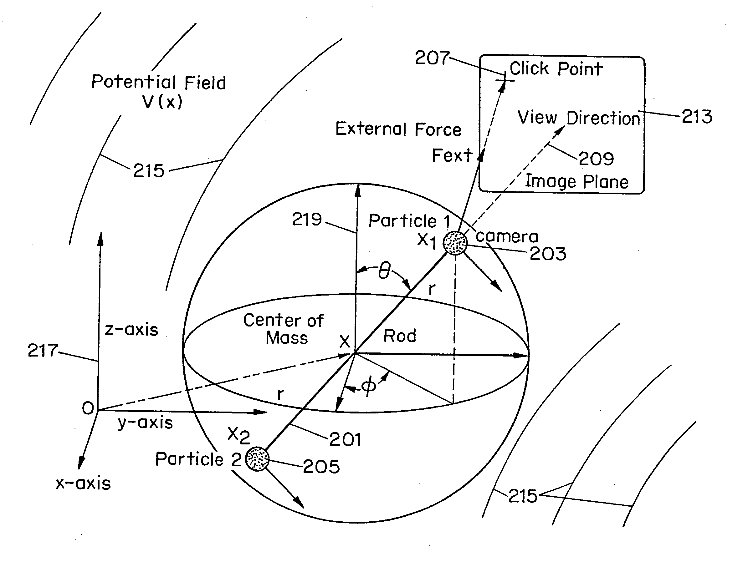
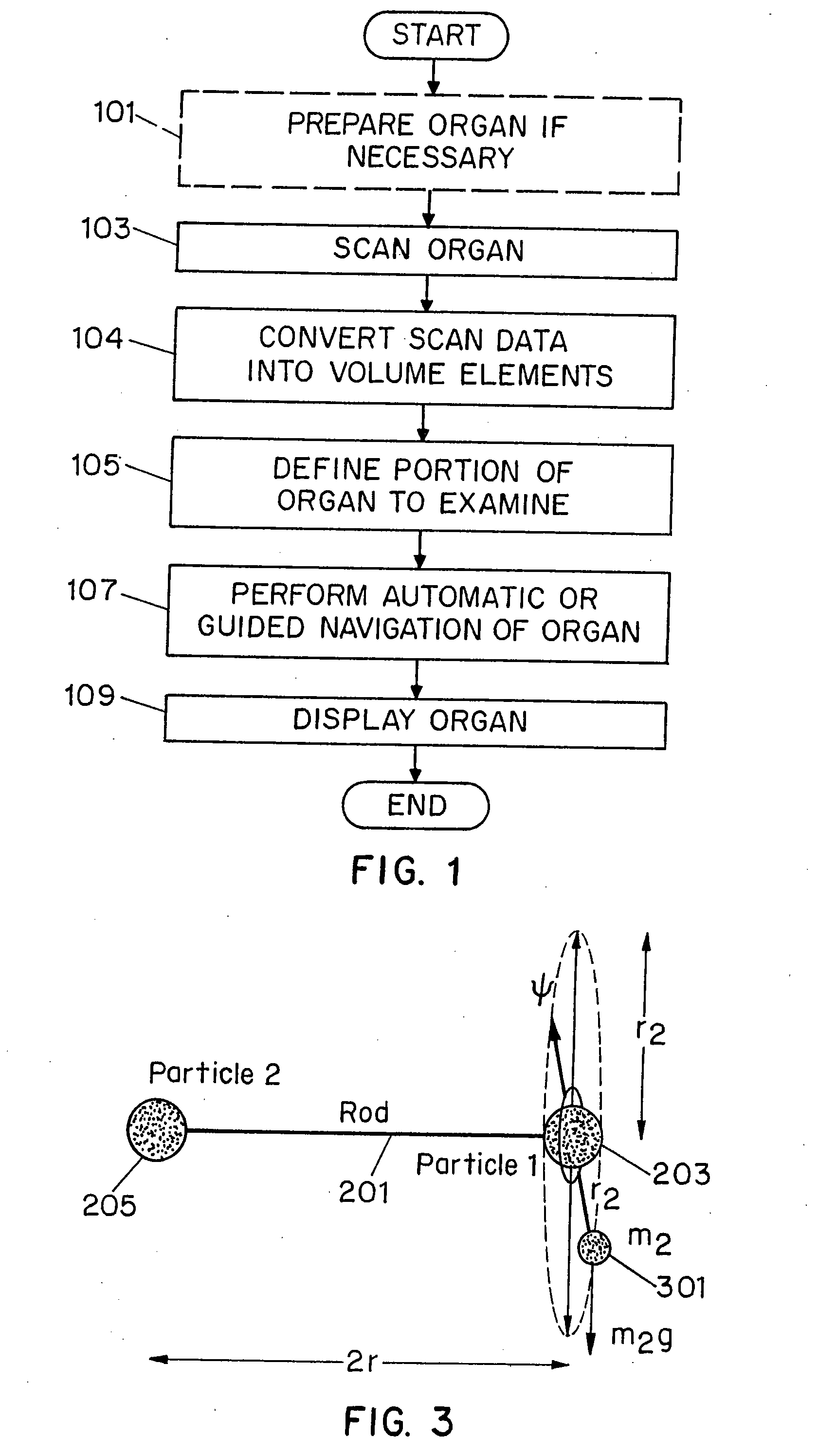
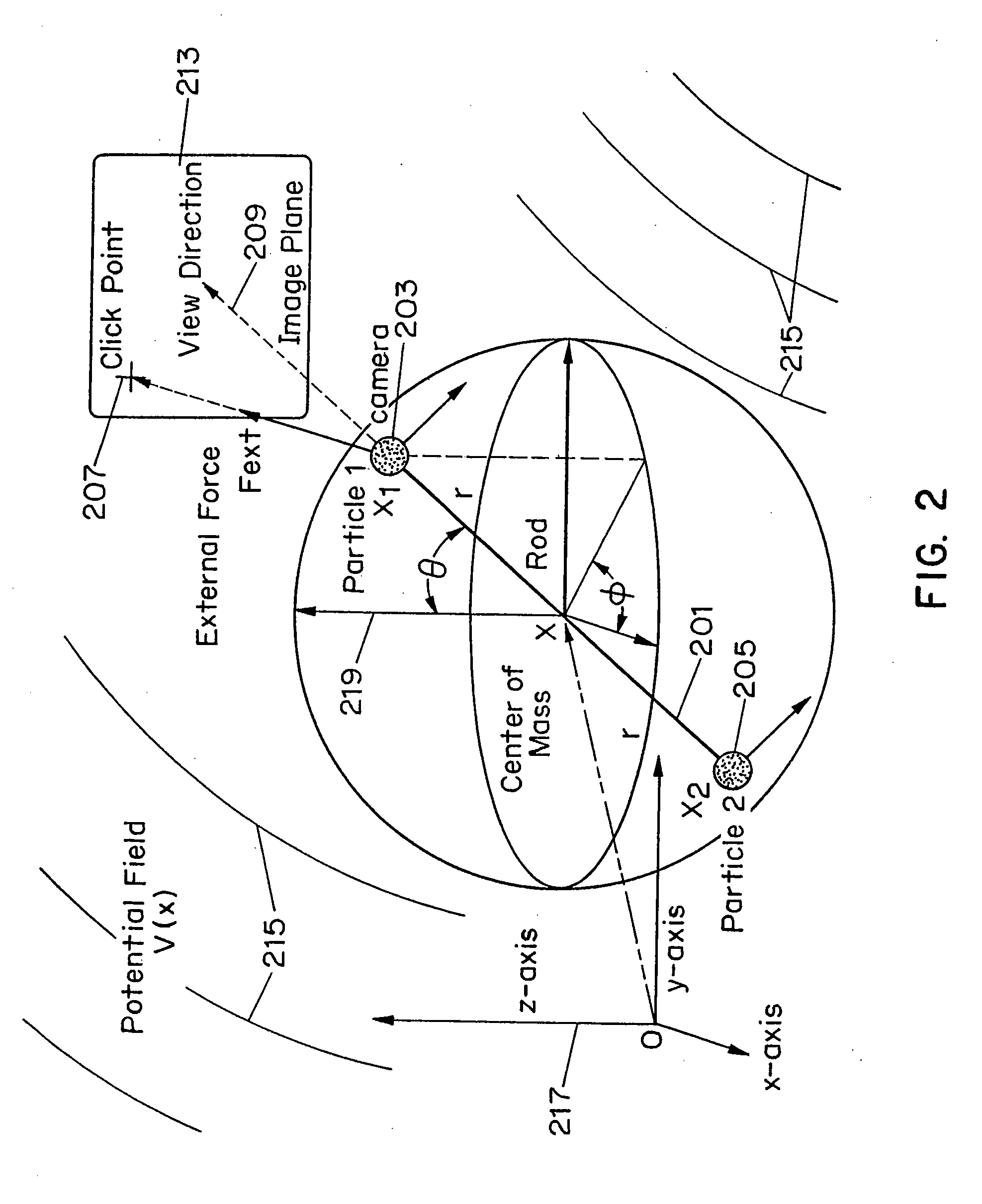
[0064]FIG. 1 illustrates the steps necessary to perform a virtual colonoscopy using volume visualization techniques. Step 101 prepares the colon to be scanned in order to be viewed for examination if required by either the doctor or the particular scanning instrument. This preparation could include cleansing the colon with a “cocktail” or liquid which enters the colon after being orally ingested and passed through the ...


PUM


Abstract

Methods for generating a three-dimensional visualization image of an object, such as an internal organ, using volume visualization techniques are provided. The techniques include a multi-scan imaging method; a multi-resolution imaging method; and a method for generating a skeleton of a complex three dimension object. The applications include virtual cystoscopy, virtual laryngoscopy, virtual angiography, among others.

Description

[0001] This application is a continuation-in-part of U.S. patent application, Ser. No. 09 / 493,559, filed on Jan. 28, 2000, entitled “System And Method For Performing a Three-dimensional Virtual Examination, Navigation and Visualization” and is a continuation-in-part of U.S. patent application, Ser. No. 09 / 343,012, filed on Jun. 29, 1999, entitled “System And Method For Performing a Three-dimensional Virtual Segmentation And Examination,” both of which are a continuation in part of U.S. Pat. No. 5,971,767, entitled “System and Method for Performing a Three Dimensional Virtual Examination.”TECHNICAL FIELD [0002] The present invention relates to a system and method for performing a volume based three-dimensional virtual examination, and more particularly relates to a system which offers enhanced visualization and navigation properties. BACKGROUND OF THE INVENTION [0003] Colon cancer continues to be a major cause of death throughout the world. Early detection of cancerous growths, which...

Claims


Application Information

Patent Timeline
Patent Type & Authority Applications(United States)
IPC IPC(8): G06T17/00
CPCG06T7/0012G06T7/0081G06T7/604G06T15/08G06T17/00G06T19/00G06T2207/10081G06T2207/20044G06T2207/20064G06T2207/20148G06T2207/30028G06T2207/30101G06T7/11G06T7/64G06T7/136
Inventor KAUFMAN, ARIE E.LIANG, ZHENGRONGWAX, MARK R.WAN, MINGCHEN, DONGQINGLI, BIN
Owner THE RES FOUND OF STATE UNIV OF NEW YORK
Learn More