System and method for airway detection

a technology of airway detection and computed tomography, which is applied in the direction of image enhancement, image analysis, electric discharge tubes, etc., can solve the problems of difficult detection of smaller airways using conventional methods, difficult detection of airways and airway walls accurately, and difficult detection of smaller airways. , to achieve the effect of accurately re-centering manual click points and accurate boundary of segmented airways

Active Publication Date: 2007-05-31
SIEMENS MEDICAL SOLUTIONS USA INC
View PDF6 Cites 9 Cited by
  • Summary
  • Abstract
  • Description
  • Claims
  • Application Information

AI Technical Summary

Benefits of technology

[0006] The present invention is directed to a system and method for detecting airways in 3D lung image data, such as CT lung image datasets. The present invention introduces operators that give high responses to airway walls, as well as filters based on these operators to detect the airways. These filters are based on multiple first and second order derivatives of the 3D lung image data and can be used in combination to detect airways. The present invention can be used as a basis for airway segmentation or quantitative analysis. For example, the present invention can be used to provide a more accurate boundary of a segmented airway, or to accurately re-center manual click points in semi-automatic segmentation methods.

Problems solved by technology

However, detection of the smaller airways using the conventional methods is difficult due to partial volume effects and noise.
Thus, it is difficult to accurately detect airways and airway walls.
Detection of smaller airways can also be difficult due to reconstruction artifacts and noise common in CT scanning.

Method used


Image

  • System and method for airway detection
  • System and method for airway detection
  • System and method for airway detection

Examples


Embodiment Construction

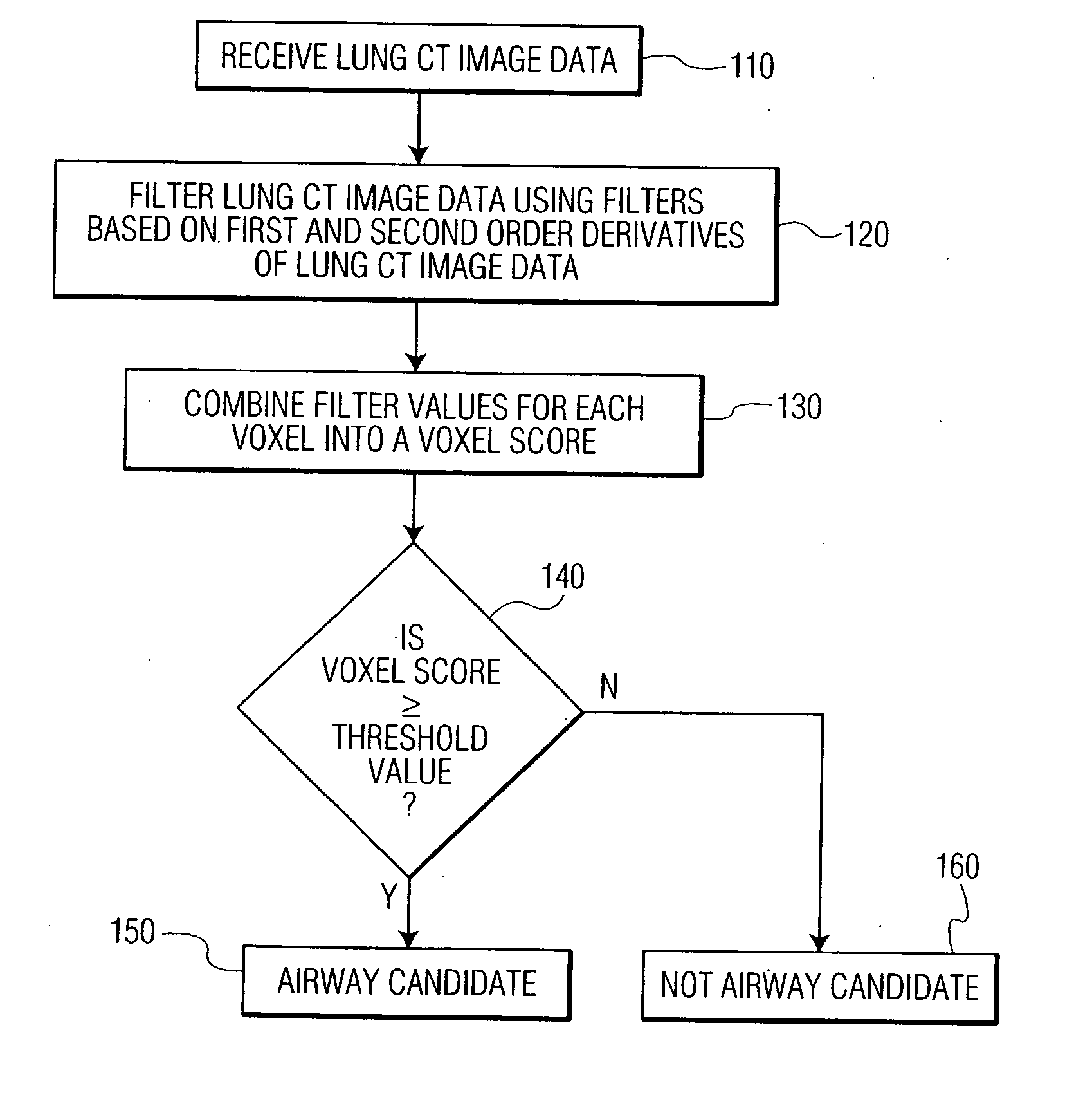
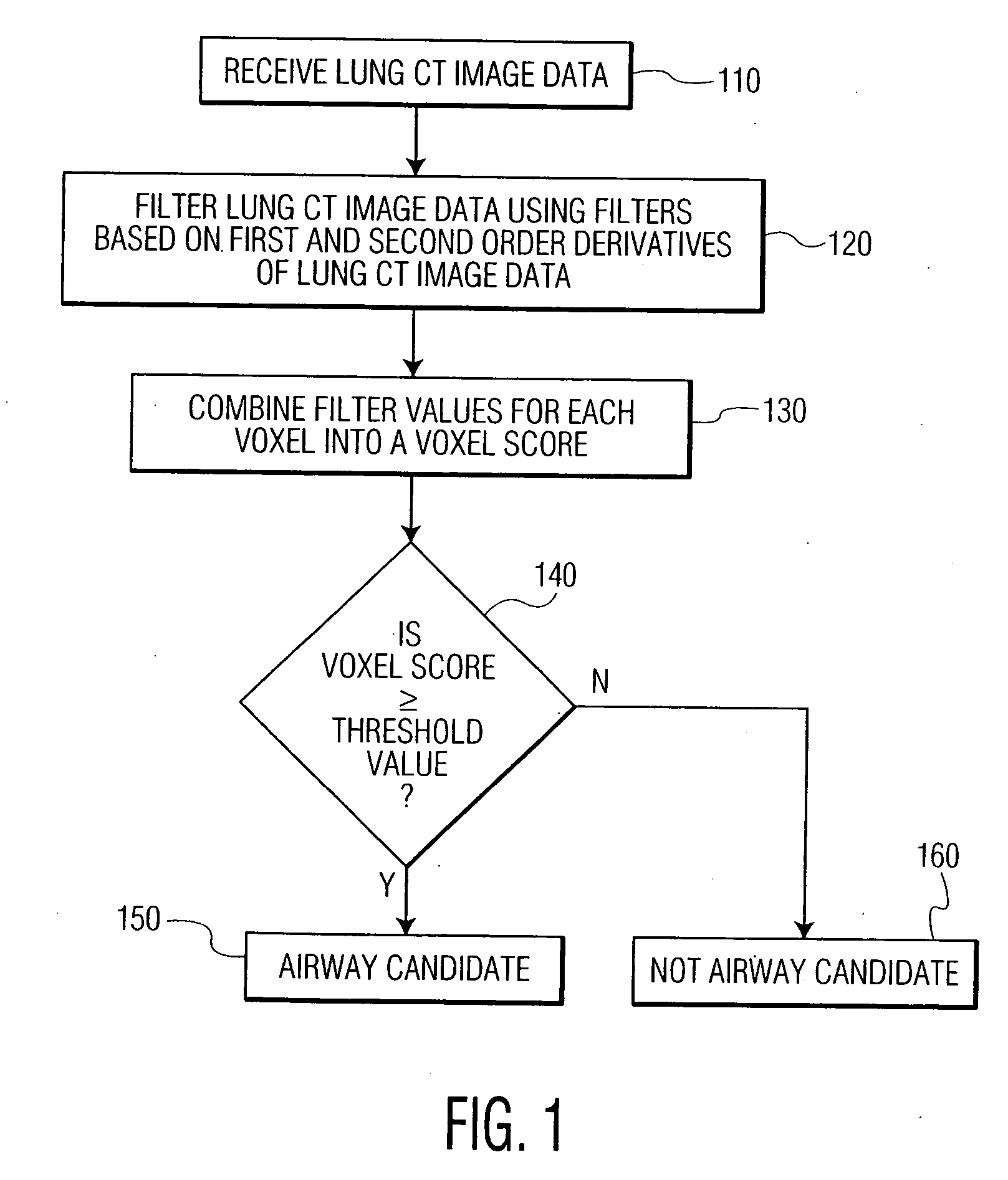
[0012]FIG. 1 illustrates a method for determining airway candidates in CT image data according to an embodiment of the present invention. At step 110, lung CT image data is received. The received lung CT image data represents a three dimensional image of the lungs. It is possible that the CT image data be input directly from a CT scanner. It is also possible that a previously stored CT dataset be retrieved in order to detect airways in the previously stored CT dataset. The lung CT image data is made up of a plurality of voxels, and each voxel has an intensity value corresponding to the density of the body tissue at that point.

[0013] At step 120, the lung CT image data is filtered using one or more image filters based on first and second order derivatives of the CT image data. Since airway walls contain a certain amount of curvature, different curvature filters can be used to highlight the airway wails in the CT image data. For example, one or more of the following filters can be im...


PUM


Abstract

A method and system for detecting airways in 3D lung image data is disclosed. The 3D lung image data is filtered using one or more filters based on first and second order derivatives of the CT image data. Each filter calculates a value for each voxel of the 3D lung image data, and the values from all of the filters are combined to determine a voxel score for each voxel. If the voxel score for a voxel is greater than or equal to a threshold value, the voxel is considered an airway candidate.

Description

[0001] This application claims the benefit of U.S. Provisional Application No. 60 / 740,416 filed Nov. 29, 2005, the disclosure of which is herein incorporated by reference.BACKGROUND OF THE INVENTION [0002] The present invention relates to detecting airways in 3-dimensional medical images of the lungs, and more particularly to a system and method for detecting airways in computed tomography (CT) lung images using filters based on first and second order derivatives of the CT lung images. [0003] Computed tomography (CT) is a medical imaging method whereby digital geometry processing is used to generate a three-dimensional image of the internal features of a patient from X-ray beams. Such CT imaging results in CT volume data which is a virtual representation of internal anatomical features of a patient. The CT volume data consists of multiple slices, or two-dimensional images, that can be combined to generate a three dimensional image, CT imaging is particularly useful because it can sh...

Claims


Application Information

Patent Timeline
Patent Type & Authority Applications(United States)
IPC IPC(8): H01J35/00
CPCG06T7/0012G06T7/0083G06T2207/10081G06T2207/30061G06T7/12
Inventor KIRALY, ATILLA PETERODRY, BENJAMINCOENEN, BJOERN E.NOVAK, CAROL L.
Owner SIEMENS MEDICAL SOLUTIONS USA INC
Why Eureka
Try Eureka