Bioresorbable Inflatable Devices, Incision Tool And Method For Tissue Expansion And Tissue Regeneration

a bioresorbable, inflatable technology, applied in medical devices, diagnostics, applications, etc., can solve the problems of inability to receive bridges, implants, dentures, and other problems, and achieve the effect of reducing the change in the shape of the inflatable elemen

Active Publication Date: 2007-07-05
KARMON BEN ZION
View PDF34 Cites 58 Cited by
  • Summary
  • Abstract
  • Description
  • Claims
  • Application Information

AI Technical Summary

Benefits of technology

[0127] The insertion of the device can be through a small incision to a tunnel so all the process is done with almost no surgery. Through this incision the tunnel incision tool can be inserted before the insertion of the device. The tunnel incision tool makes shallow incision in the surrounding tissue of the tunnel. These incisions allow initial expansion of the tissue and easy insertion of the device.
[0236] According to a further feature of the present invention the inflatable element is configured to take a specific shape as the inflatable element is filled and to minimize changes in the shape of the inflatable element as a result of forces coming from outside the inflatable element.

Problems solved by technology

The absence of just one tooth can cause modifications throughout the dental arch and even prompt a possible softening (loss of insertion) which may cause the loss of other teeth.
Bone loss may finally modify the patient's appearance and, depending on the loss, may make him incapable of receiving bridges, implants or even dentures.
Although these methods of surgical reconstruction have been successfully performed, this type of operation has had drawbacks.
Exposure of the membrane leading to infection and failure of the regeneration.
All the hazards of a relative big operation in the mouth: bleeding, nerve damage, infection, pain etc.
It is difficult with this technique to place accurately the graft 2.
The surgeon is often unable to achieve the desired reconstruction of the atrophied ridge without perforating or stretching of the mucoperiosteum to the point that pressure necrosis develops.
While this expandable device expands tension is transferred to the periosteum leading to enlargement of the periosteum.
The technique is complicated.
The Schneiderian membrane can be easily torn resulting in infection of the sinus and failure of the operation.
The amount of augmentation is limited.
The Schneiderian membrane can be torn without the awareness of the surgeon resulting in filling the graft above the membrane and failure of the procedure.
The technique is complicated.
There is an intentional tear of the membrane that can cause infection and failure of procedure.
The technique requires special equipment and skill that are not familiar to the surgeons involved in dental implantology.
This type of device proved to be complex and limited by a small expansion ration.
Another self-expanding stent used stainless steel wire in a zig zag configuration which resulted in incomplete vascular contact and only partial healing of the device.
Thrombosis occurred in these early prototypes, especially when the vessel tapered, and at branch points and at low expansion ratios.
Fischell et al teaches that the thrombogenic potential of the device is reduced, through a passive methodology, but does nothing to address the biological response to the implant as a foreign body.
Moreover, no suggestion is made of a way to inhibit neointimal hyperplasia, which inevitably follows balloon catheter induced injury to arterial vessels.
Concern here, of course, is that the inside located ledge and other retaining means may inadvertently function to cause further blockage of the tube once it is installed.
The obvious concern is that the size must be precise or the tube will expand too much or too little, either damaging the vessel or escaping from the location for which it was intended.
Second, the delivery system must facilitate the deployment of the stent once located in a vessel.
Third, the stent delivery system must easily disengage from the stent after the stent is deployed.
Sometimes this reduced the effectiveness of the therapeutic agent, particularly when the area of treatment was displaced from the second stent.
However, it is still a fact that each insertion and extraction risks further damage to afflicted areas and damage to otherwise unaffected areas through which the instruments pass and can add to patient trauma.
Moreover, insertion and withdrawal of additional instruments in sequence increases the time of the physician, staff and medical facility, and the cost of multiple instruments.
None of the prior art is enabling the widening of the vessel without temporarily blocking the passage through the vessel.
None of the prior art enables gradually widening of the vessel over some days therefor limiting the risk of rupture.
None of the prior art enables safely widening the vessel beyond the final desired diameter to compensate for future restenosis.

Method used


Image

  • Bioresorbable Inflatable Devices, Incision Tool And Method For Tissue Expansion And Tissue Regeneration
  • Bioresorbable Inflatable Devices, Incision Tool And Method For Tissue Expansion And Tissue Regeneration
  • Bioresorbable Inflatable Devices, Incision Tool And Method For Tissue Expansion And Tissue Regeneration

Examples


Embodiment Construction

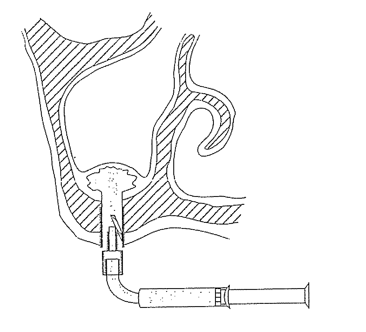
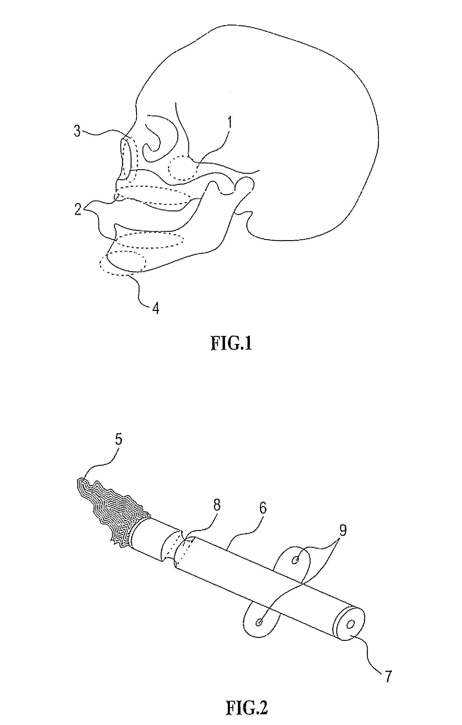
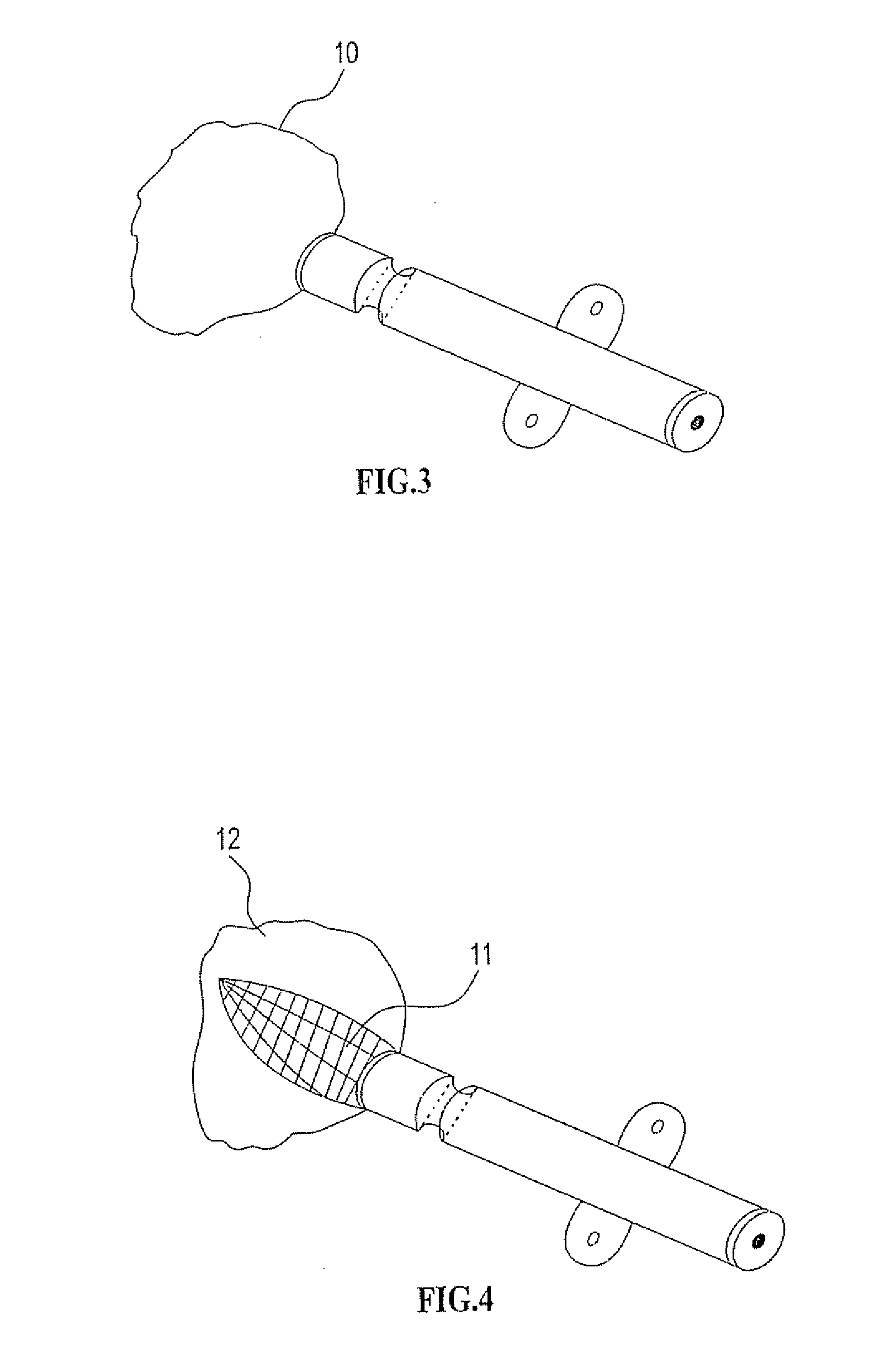
[0310] As mentioned further above there are many implementations of the invention in different tissues and organs. The following description will focus on embodiments in three fields in order to understand the principles of the device and method. The first is bone augmentation in the jaws the second is vessel widening using a bioresorbable stent, the third is fixating bone fragments. The same principles should be used in other tissues and organs.

[0311] Before turning to the features of the present invention in more detail, it will be useful to clarify certain terminology as will be used herein in the description and claims. Specifically, it should be noted that the present invention is useful in a wide range of applications in which living tissue is to be expanded, stretched or displaced. The term “living tissue” is used herein to refer to any living tissue including, but not limited to, an organ, tube, vessel, cavity, bone cavity or membrane, and interfaces between any two or more...


PUM

PropertyMeasurementUnit
widthaaaaaaaaaa
heightaaaaaaaaaa
depthaaaaaaaaaa

Abstract

Bioresorbable inflatable devices and tunnel incision tool and methods for treating and enlarging a tissue or an organ or a tube or a vessel or a cavity. The device is composed of a hollow expanding pouch made of a resorbable material or a perforated material that can be attached to a filling element. The pouch can be filled with a biocompatible materials, one or more times in few days interval, after the insertion of the device. While filling the pouch every few days the tissue expands and the filling material if it is bioactive start to function. The tunnel incision tool composed of a little blade that emerges from the surface of the tool in order to make shallow incisions in the surrounding tissue therefore enabling easy expansion of the tissue. This device and method can be used for example for: horizontal and vertical bone augmentation in the jaws and the tunnel incision tool is used to make shallow incisions in the periosteum when using the tunnel technique, sinus augmentation when the device is placed beneath the Schneiderian tissue, vessels widening if the pouch become a stent, fixating bone fractures etc.

Description

[0001] This application is a Continuation of U.S. patent application Ser. No. 10 / 291,477 filed Nov. 12, 2002, which is a CIP of International Application No. PCT / IL2001 / 00408, filed on May 9, 2001 and claiming priority from U.S. patent application Ser. No. 09 / 567,471 filed May 9, 2000FIELD AND BACKGROUND OF THE INVENTION [0002] The present invention relates to an improved methods and devices for treating and healing a tissue deficiency in a living human or animal body. The method and the device combine a mechanical action and a biological action. [0003] For example, the present invention can be used for guided bone regeneration in the jaws as part of dental treatment with dental implants. [0004] The present invention consists of an expansion device based on a bioresorbable film or a perforated film and a method for tissue regeneration. In order to clarify the principles of the present invention the following description will focus on three implementations: bone regeneration in the j...

Claims


Application Information

Patent Timeline
Patent Type & Authority Applications(United States)
IPC IPC(8): A61F2/28A61B17/00A61B17/58A61B17/66A61B17/68A61B17/70A61B17/74A61B17/88A61B19/00A61C8/00A61F2/00A61F2/02A61F2/30A61F2/36A61F2/44A61F2/46
CPCA61B17/58Y10S623/908A61B17/68A61B17/70A61B17/7097A61B17/7098A61B17/742A61B17/8802A61B17/8811A61B17/8855A61B19/54A61B2017/00004A61B2017/00557A61C8/0006A61C8/0009A61C8/0092A61F2/30723A61F2/30724A61F2/30734A61F2/36A61F2/3662A61F2/3676A61F2/44A61F2/4601A61F2002/2828A61F2002/2832A61F2002/30014A61F2002/30062A61F2002/3008A61F2002/30581A61F2002/30738A61F2002/30919A61F2002/30932A61F2002/3631A61F2002/4631A61F2210/0004A61F2250/0018A61F2250/0098A61B17/666A61B90/39A61M31/002A61M2205/0211A61M2210/0681
Inventor KARMON, BEN-ZION
Owner KARMON BEN ZION
Why Eureka
Try Eureka