Ultrasound probe for paracentesis and ultrasound diagnostic apparatus

a paracentesis and ultrasound technology, applied in the field of ultrasound diagnostic equipment and paracentesis ultrasound probe, can solve the problems of inability to accurately position the biopsy needle, and the method in question is not suitable for other ultrasound inspections

Inactive Publication Date: 2007-07-19
GE MEDICAL SYST GLOBAL TECH CO LLC
View PDF12 Cites 32 Cited by
  • Summary
  • Abstract
  • Description
  • Claims
  • Application Information

AI Technical Summary

Problems solved by technology

However, it is impossible to collect image data of the portion positioned just under the biopsy needle guide hole formed centrally of the probe surface, so there occurs a dropout portion (dead angle) of image near the central part of the upper side of the sectional image and thus the method in question is not suitable ...

Method used


Image

  • Ultrasound probe for paracentesis and ultrasound diagnostic apparatus
  • Ultrasound probe for paracentesis and ultrasound diagnostic apparatus
  • Ultrasound probe for paracentesis and ultrasound diagnostic apparatus

Examples


Embodiment Construction

[0023] An ultrasound probe for paracentesis according to an embodiment of the present invention will be described hereinunder concretely with reference to the drawings.

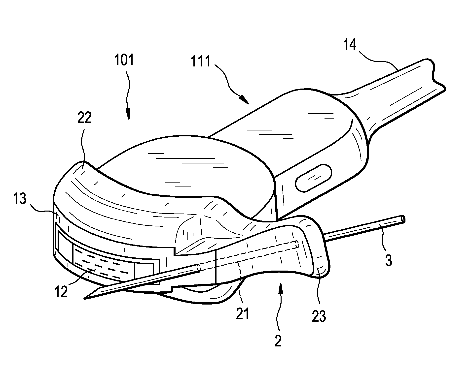
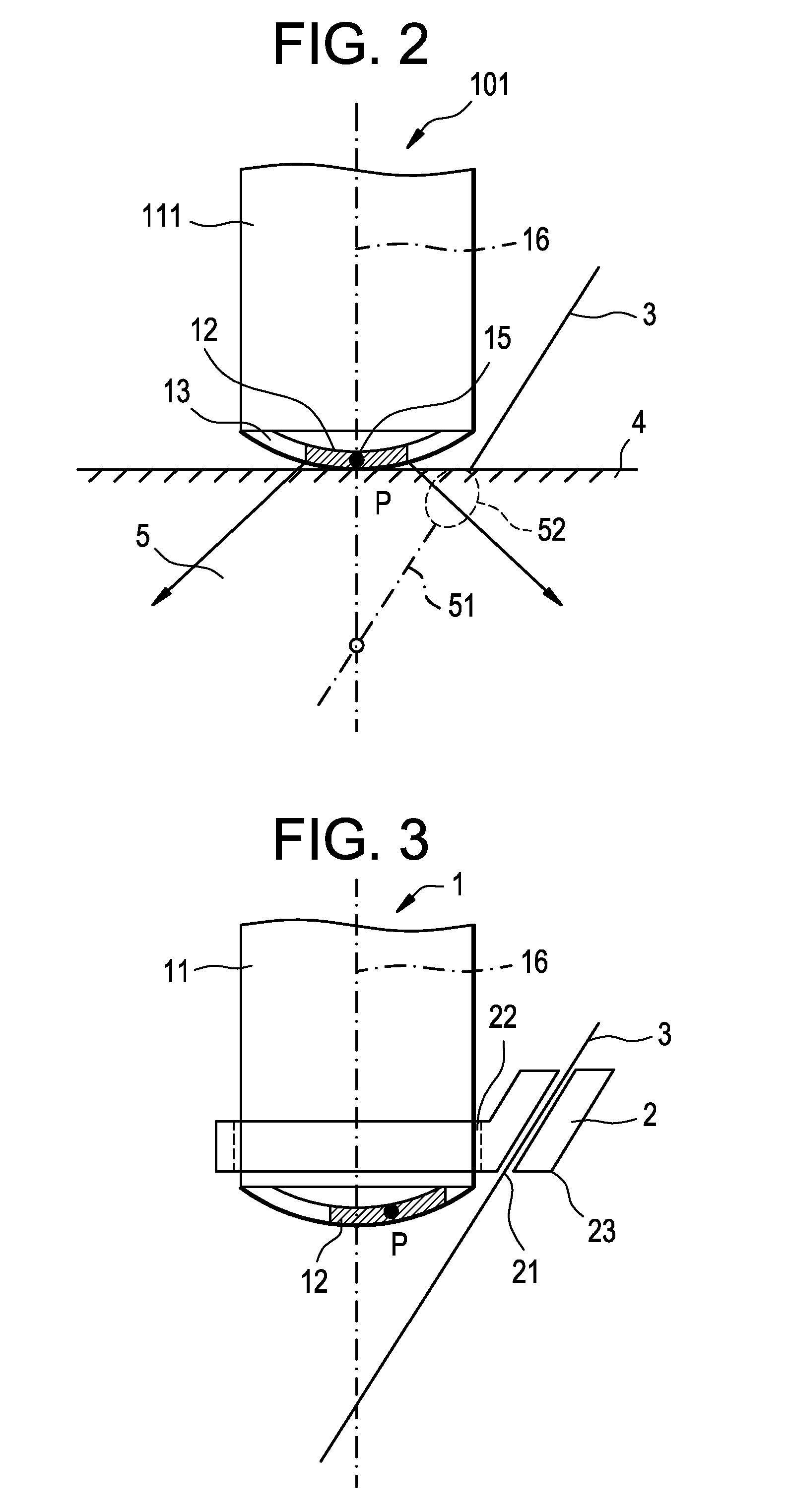
[0024]FIG. 3 is a sectional view of an ultrasound wave-generating tip portion of the ultrasound probe 1 for paracentesis, showing an assembled state of a probe body 11 and an adapter 2. FIG. 3 is a sectional view of the tip portion of the ultrasound probe 1 for paracentesis, but an entire perspective view of the probe is omitted because it is common to the conventional ultrasound probe shown in FIG. 1.

[0025] As shown in FIG. 3, the adapter 2 for biopsy needle for the ultrasound probe is attached to the ultrasound probe 1 detachably. The adapter 2 is made up of an attaching / detaching portion 22 capable of being attached to and detached from the probe body 11 and a prescribing portion 23 for prescribing a biopsy needle 3 onto an ultrasound scanning line by the ultrasound probe 1. The attaching / detaching portion 22 of ...


PUM


Abstract

An ultrasound probe for paracentesis with a biopsy needle attached to a probe body, which permits an operator to insert the biopsy needle into an exact position of a patient, is to be provided. An ultrasound probe for paracentesis comprising a probe body and a biopsy needle attached to the probe body, wherein the center of an acoustic elements array provided in the probe body is offset toward the biopsy needle attached to the probe body with respect to a central vertical line of the probe body.

Description

BACKGROUND OF THE INVENTION [0001] The present invention relates to an ultrasound probe for paracentesis and an ultrasound diagnostic apparatus having the ultrasound probe for paracentesis. [0002] Paracentesis has been conducted wherein a biopsy needle such as an injection needle is inserted into a living body to sample a tumor tissue for example or for local administration of a drug. For avoiding damage of a blood vessel which would cause a large quantity of bleeding or for positive paracentesis of a tumor tissue for example, the paracentesis is usually performed while making reference to a sectional image formed by an ultrasound diagnostic apparatus. [0003] As methods for performing the paracentesis while being guided by the ultrasound diagnostic apparatus there are known a method wherein an ultrasound probe for paracentesis is used and a biopsy needle is inserted into a living body through a guide hole formed centrally of a probe surface (serving not only as an ultrasound irradia...

Claims


Application Information

Patent Timeline
IPC IPC(8): A61B8/14
CPCA61B8/0833A61B8/0841A61B2019/5276A61B17/3403A61B2017/3413A61B8/14A61B2090/378A61B8/00
Inventor NOZAKI, MITSUHIRO
Owner GE MEDICAL SYST GLOBAL TECH CO LLC
Why Eureka
Try Eureka