Systems and Methods for Simulation of Organ Dynamics

a system and organ technology, applied in the field of systems and methods for simulation of organ dynamics, can solve the problems of inability to provide more advanced visualization paradigms, inability to provide discrete images, and inability to provide predictive components regarding the motion of specific organs

Inactive Publication Date: 2007-10-04
UNIV OF CENT FLORIDA RES FOUND INC
View PDF16 Cites 105 Cited by
  • Summary
  • Abstract
  • Description
  • Claims
  • Application Information

AI Technical Summary

Problems solved by technology

Despite those benefits, inherent drawbacks exist.
Additionally, the discrete nature of the images does not permit more advanced visualization paradigms, such as a virtual reality, to enable the development of advan

Method used


Image

  • Systems and Methods for Simulation of Organ Dynamics
  • Systems and Methods for Simulation of Organ Dynamics
  • Systems and Methods for Simulation of Organ Dynamics

Examples


Embodiment Construction

[0016] As described above, there are various limitations to current medical imaging techniques, one such limitation being the inability to predict the dynamic motion of an organ. Disclosed herein, however, are systems and methods that can be used to real-time simulate organ dynamics in three-dimensions.

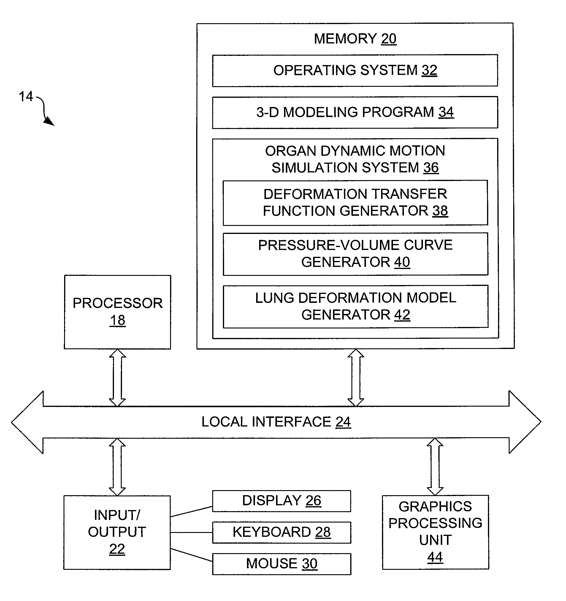
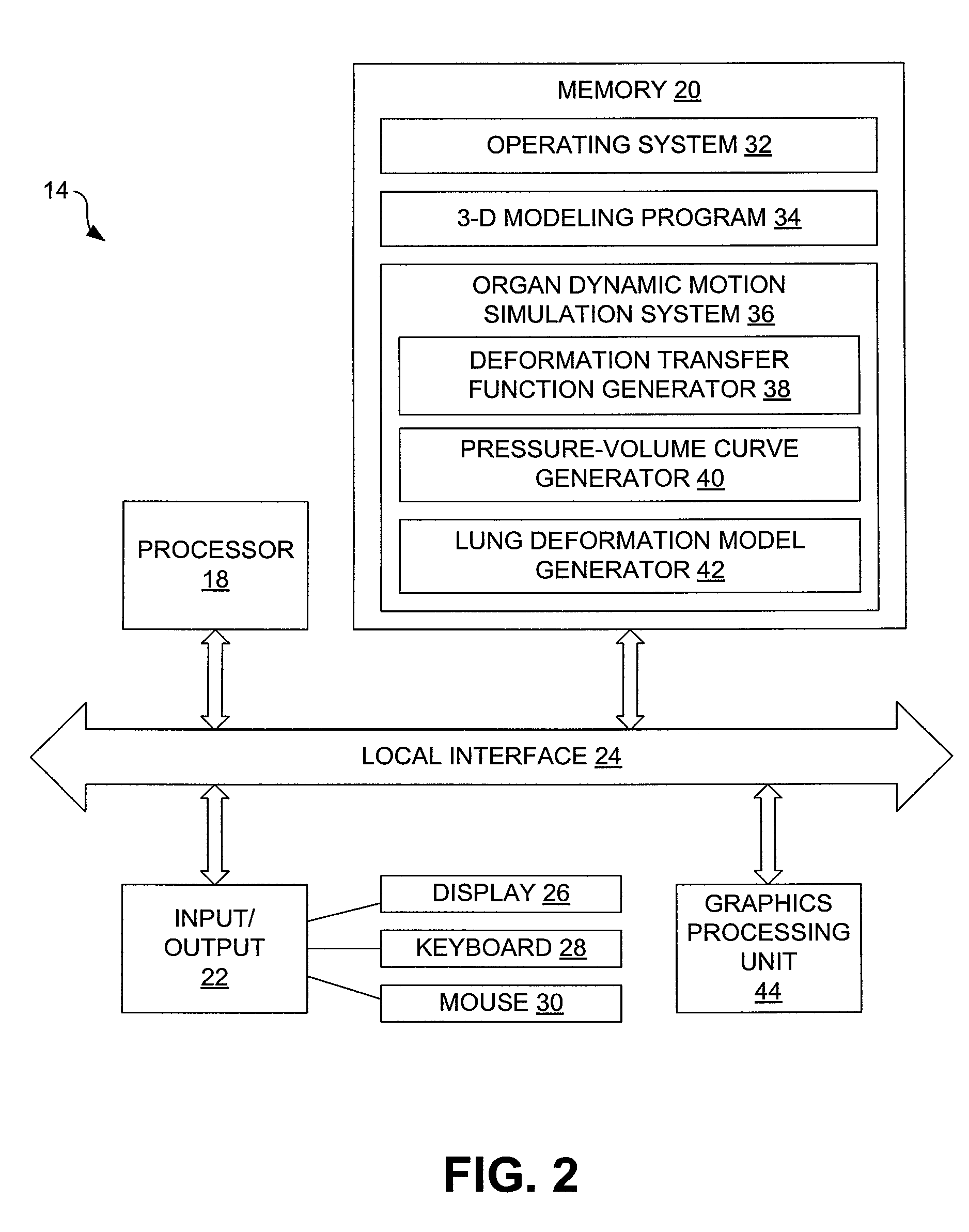
[0017] In some embodiments, the systems and methods can be used to simulate dynamic motion of human lungs. To create such a simulation, images of the lungs of a subject are captured during breathing and are used to generate a three-dimensional lung model. As described in greater detail below, the lung model can then be used to generate a deformation transfer function and a pressure-volume curve. Once the deformation transfer function and the pressure-volume curve are generated, they can be used to generate dynamic lung models for various instances of time.

[0018] Although simulation of lung dynamics is described with specificity in this disclosure, it is to be appreciated that the di...


PUM


Abstract

In one embodiment, a method for simulating organ dynamics includes generating a sequence of three-dimensional models of an organ during different stages of observed dynamic motion, generating a deformation transfer function from the sequence of three-dimensional models, generating a pressure-volume curve from the sequence of three-dimensional models, and generating an organ deformation model that simulates dynamic motion of the organ.

Description

CROSS-REFERENCE TO RELATED APPLICATION [0001] This application claims priority to copending U.S. provisional application Ser. No. 60 / 773,486 entitled “OPTICAL APPARATUSES AND METHODS,” which was filed Feb. 15, 2006, and which is entirely incorporated herein by reference.BACKGROUND [0002] Medical imaging provides numerous benefits in relation to patient diagnosis and treatment. Despite those benefits, inherent drawbacks exist. For example, in typical cases, only discrete sets of still images or highly isolated real-time images are generated that are dependent of the position of a transducer or other image gathering device. Additionally, the discrete nature of the images does not permit more advanced visualization paradigms, such as a virtual reality, to enable the development of advanced patient education tools and surgical simulators. Further, current imaging technologies do not provide a predictive component regarding the motion of a specific organ, such as a lung during the patien...

Claims


Application Information

Patent Timeline
IPC IPC(8): G09B23/28
CPCG09B23/28
Inventor SANTHANAM, ANAND P.ROLLAND, JANNICKFIDOPIASTIS, CALI M.
Owner UNIV OF CENT FLORIDA RES FOUND INC
Why Eureka
Try Eureka