Medical imaging diagnosis supporting apparatus and image diagnosis supporting method

a technology for medical imaging and supporting equipment, applied in the field of imaging diagnosis supporting equipment, can solve the problems of conventional methods, inability to use images, and inability to give to other institutes

Active Publication Date: 2007-10-11
TOSHIBA MEDICAL SYST CORP
View PDF3 Cites 21 Cited by
  • Summary
  • Abstract
  • Description
  • Claims
  • Application Information

AI Technical Summary

Benefits of technology

[0008]The present invention has been achieved in consideration of the above-described circumstances and an object of the invention is to provide a medical imaging diagnosis supporting apparatus and an imaging diagnosis supporting method of generating preferable introduction information of a patient from an introducing-side medical institution to another medical institution from the medical viewpoints and the like, and capable of determining whether various information is provided from the introducing-side medical institution to another medical institution or not, an information providing method, and content of introduction information from the viewpoint of capability of network environment and security against information leakage.

Problems solved by technology

The conventional method, however, has the following problems.
Even if a patient is introduced with images, there is a case that the another institute cannot use the images due incompatibility of an examination apparatus or the model.
When there are many images, all of even key images cannot be stored in some media, so that they cannot be given to the another institute.
Further, when medical images and the like as personal information are recorded on a medium, a problem on security such as information leakage may occur.

Method used


Image

  • Medical imaging diagnosis supporting apparatus and image diagnosis supporting method
  • Medical imaging diagnosis supporting apparatus and image diagnosis supporting method
  • Medical imaging diagnosis supporting apparatus and image diagnosis supporting method

Examples


first embodiment

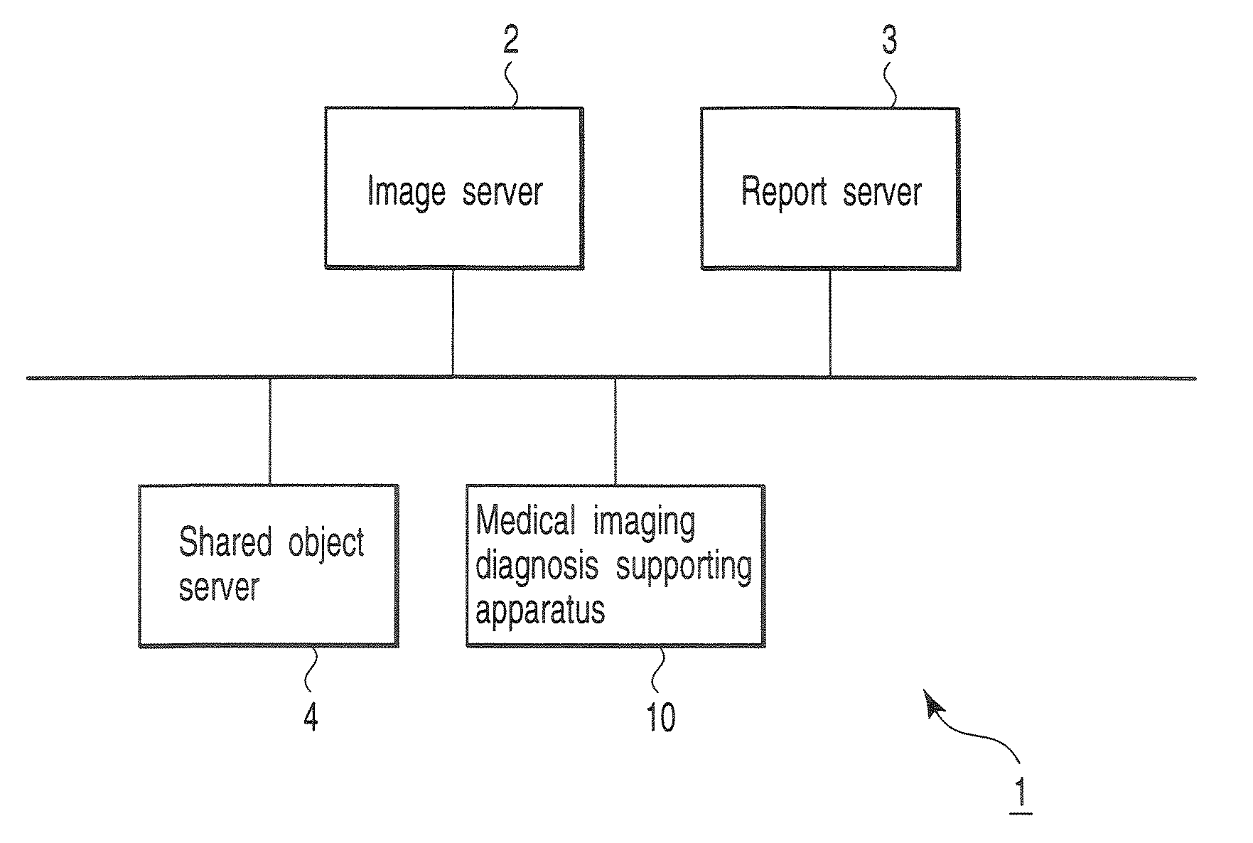
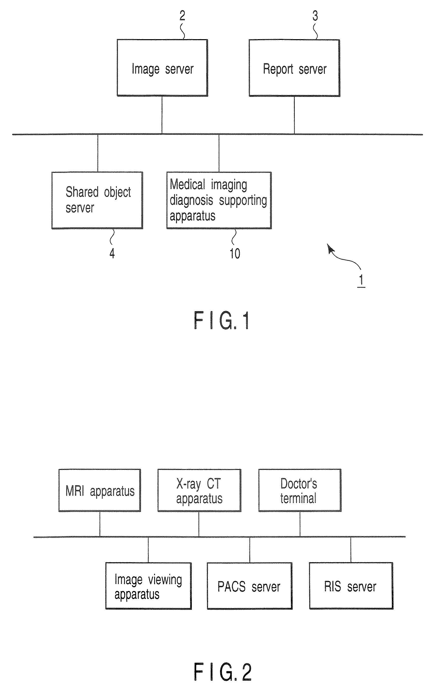
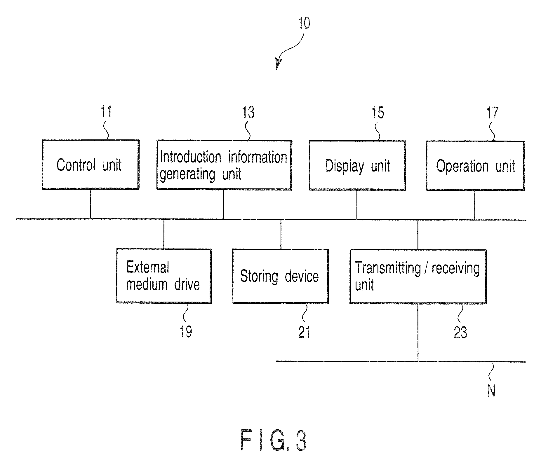
[0024]FIG. 1 is a block diagram showing the configuration of a medical diagnostic imaging support system 1 according to the present embodiment. As shown in the diagram, the medical diagnostic imaging support system 1 has an image server 2, a report server 3, a shared object server 4, and a medical imaging diagnosis supporting apparatus 10.

[0025]The image server 2 manages images generated by various medical diagnostic imaging apparatuses, images generated by post-process by an image viewing apparatus 7, and the like by using series IDs, patient IDs, dedicated management IDs, and the like. A series is a concept for managing various information by time (when information is generated), space (where information is generated), and an clinical characteristic of information (clinical meaning). Therefore, for example, a plurality of CT images captured by a single scan in an examination using an X-ray CT apparatus are information belonging to the same series. A series ID is an identification ...

second embodiment

[0083]A second embodiment of the present invention will be described. A medical imaging diagnosis supporting apparatus according to the second embodiment stores, as introduction information, not image data and the like itself to an external medium but address information for accessing image data and the like to be provided to another institute via a network.

[0084]FIG. 7 is a sequence chart showing the flow of introduction information generating process according to the second embodiment. As shown in the diagram, in steps S11 to S14, processes similar to those of the steps S1 to S4 shown in FIG. 6 are executed.

[0085]Next, the introduction information generating unit 13 of the medical imaging diagnosis supporting apparatus 10 transmits a request for obtaining address information for accessing various kinds of content (that is, a selected key image, report information, and a shared object having the same series ID as that of the key image) to the various servers such as the image serve...

third embodiment

[0092]For example, in the first embodiment, when an external medium does not have enough capacity to store all of content data such as key image data, a shared object, and the like, preferably, part of data is provided to another institute to which a patient is introduced via a network as necessary. In this case or also in the second embodiment, it is preferable to regulate kinds of data to be obtained by another institute from an introducing-side institute via a network from the viewpoint of security against information leakage and from the viewpoint of the network environment (mainly, line speed) of the another institute.

[0093]The medical diagnostic imaging support system of the third embodiment automatically determines content to be included in introduction information, whether or not various kinds of content are provided to another institute to which a patient is introduced, and a providing method in accordance with the security level, line speed, and the like of the another ins...


PUM


Abstract

By selecting at least one of an image and a report to be provided for an institute to which a patient is introduced, an object having the same series ID as that of the image or report is automatically obtained. Medical information including the object is generated and stored in an external medium. By selecting an image or the like to be provided to the institute to which a patient is introduced, address information of the selected image or the like and address information of an object or the like having the same series ID as that of the image or the like is automatically obtained. Medical information including the address information is generated and stored in an external medium.

Description

CROSS-REFERENCE TO RELATED APPLICATIONS[0001]This application is based upon and claims the benefit of priority from prior Japanese Patent Application No. 2006-061499, filed Mar. 7, 2006, the entire contents of which are incorporated herein by reference.BACKGROUND OF THE INVENTION[0002]1. Field of the Invention[0003]The present invention relates to an imaging diagnosis supporting system and, more particularly, to a medical imaging diagnosis supporting apparatus and an imaging diagnosis supporting method used when data is transmitted / received among a plurality of apparatuses.[0004]2. Description of the Related Art[0005]In recent years, a medical activity is finely divided. For example, image diagnosis is divided into works of generation of an examination order of a patient, acquisition of diagnostic images based on the generated order, reading of the acquired diagnostic images and generation of a report, and explanation of the diagnosis result and treatment course on the basis of the ...

Claims


Application Information

Patent Timeline
Patent Type & Authority Applications(United States)
IPC IPC(8): G06Q10/00G06F19/00A61B5/00A61B6/03G16H10/60G16H15/00G16H30/20G16H50/20
CPCG06F19/321G06Q50/24G06Q50/22G16H50/20G16H30/20G16H15/00
Inventor MASUZAWA, TAKASHIMATSUE, KENJINIWA, KENICHI
Owner TOSHIBA MEDICAL SYST CORP
Why Eureka
Try Eureka