
Method and Apparatus to Detect Biocontamination in an Insufflator for use in Endoscopy

a biocontamination and endoscopy technology, applied in the field of endoscopy equipment, can solve the problems of putting patients at greater risk, unable to determine whether the backflow contains biocontamination, and complicating the cleaning and sterilization of equipmen

Active Publication Date: 2007-10-18
STRYKER CORP
View PDF14 Cites 11 Cited by
  • Summary
  • Abstract
  • Description
  • Claims
  • Application Information

AI Technical Summary

Problems solved by technology

If biocontamination reaches the insufflator, contamination of the insulator can occur, which necessitates a delay in the procedure, possibly putting the patient at greater risk and, at the very least, complicating cleaning and sterilization of the equipment.
With conventional insufflators, there is no easy way to determine whether backflow contains biocontamination or merely sterile gas, other than by visual inspection.
Visual inspection, however, can be difficult and inaccurate.
Some systems use a humidity detector to detect indirectly the possible presence of backflow; however, that is not a very accurate detection method, at least because it cannot directly detect the presence of biocontamination.
Among other reasons, a humidity detector cannot reliably detect the presence of biological particulate matter independently of body fluid.
There may be other reasons why it may be undesirable to base the detection of biocontamination or backflow on humidity.
Known conventional insufflators, however, have no way of detecting this mode of biocontamination.

Method used


Image

  • Method and Apparatus to Detect Biocontamination in an Insufflator for use in Endoscopy
  • Method and Apparatus to Detect Biocontamination in an Insufflator for use in Endoscopy
  • Method and Apparatus to Detect Biocontamination in an Insufflator for use in Endoscopy

Examples


Embodiment Construction

[0012] An improved insulator for endoscopy is described. References in this specification to “an embodiment”, “one embodiment”, or the like, mean that the particular feature, structure or characteristic being described is included in at least one embodiment of the present invention. Occurrences of such phrases in this specification do not necessarily all refer to the same embodiment.

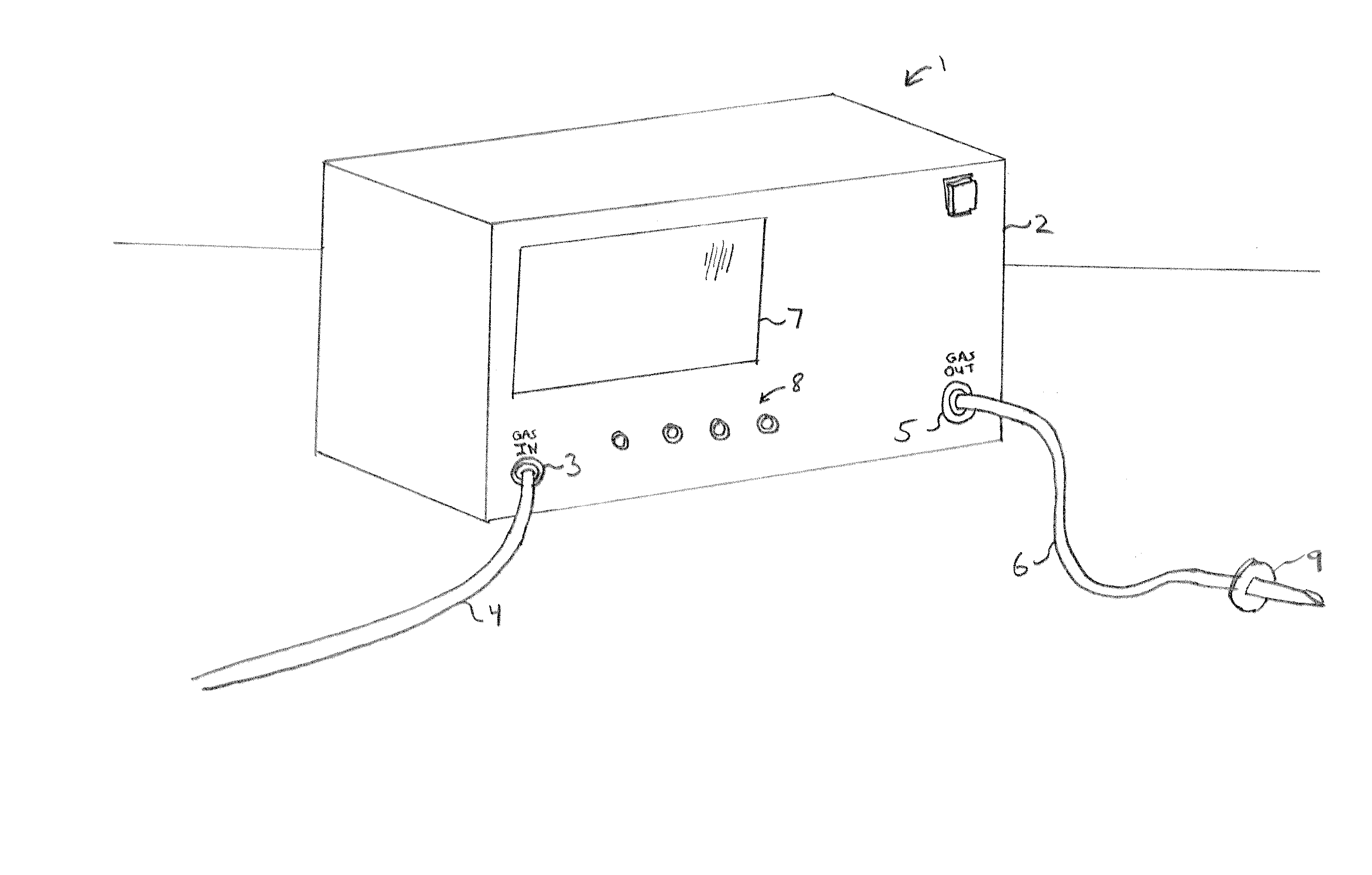
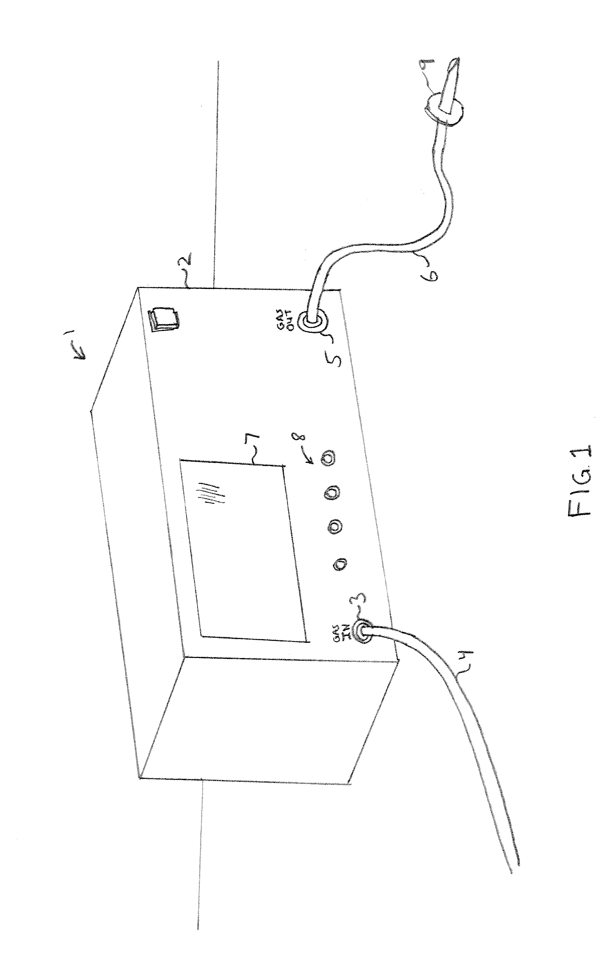
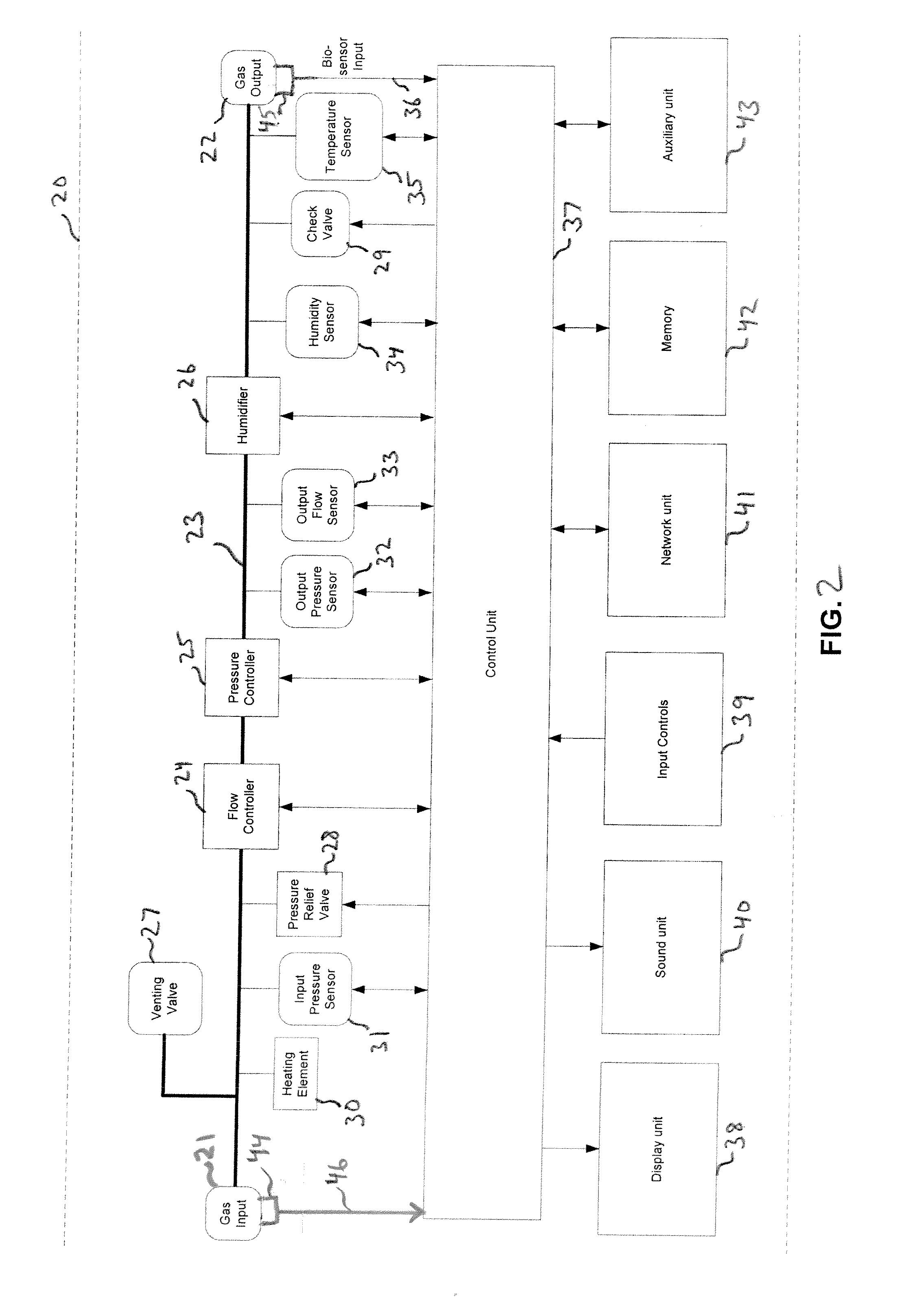
[0013] Refer now to FIG. 1, which shows an example of some of the external features of an insulator, according to certain embodiments of the invention. The insufflator 1 has a housing 2, within which are contained one or more pressure regulators and flow rate regulators and other components (not shown in FIG. 1). The housing 1 has a gas input interface 3 at which a flexible gas conduit 4 is connected to receive the gas from an external gas canister (not shown) and a gas output port 5 at which another flexible gas conduit 6 can be connected to provide gas to the patient. Conduit 6 terminates in a trocar ...


PUM


Abstract

An apparatus to provide a regulated pressurized gas for introduction into the body of a patient during a medical procedure comprises an internal flow path through which to convey the pressurized gas, a regulator in the flow path to regulate the pressurized gas, and a biosensor to detect a body substance entering or in proximity to the flow path.

Description

FIELD OF THE INVENTION [0001] At least one embodiment of the present invention pertains to equipment used for endoscopic medical procedures, and more particularly, to an advanced insulator for endoscopy. BACKGROUND [0002] Endoscopy in the medical fields allows internal structures of a patient's body to be viewed without the use of traditional, fully-invasive surgery. Endoscopy is widely used to perform many types minimally-invasive medical procedures, such as arthroscopy, laparoscopy, gastroscopy, colonoscopy, etc. A basic tool of endoscopy is the endoscope (“scope”), which contains a set of optics through which internal features of a patient's body can be viewed (typically, with the aid of a special-purpose video camera attached to the endoscope and an appropriate video monitor), when the endoscope is partially inserted into the patient's body. [0003] A supporting device commonly used in certain forms of endoscopy is an insulator. An insufflator is an electromechanical device which...

Claims


Application Information

Patent Timeline
IPC IPC(8): A61M37/00
CPCA61M13/003A61M2205/3331A61M2205/3561A61M2205/52A61M2205/362A61M2205/502A61M2205/3584
Inventor HAMEED, SALMAANMAHADIK, AMIT A.GATTANI, ABHISHEKIYER, NATRAJ
Owner STRYKER CORP
Learn More