Integrated X-Ray and Ultrasound Medical Imaging System

a medical imaging system and ultrasound technology, applied in the field of medical imaging systems, can solve the problems of “false negatives”, “false positives”, and require a second patient office visit, and achieve the effect of reducing instances, increasing sensitivity and specificity

Inactive Publication Date: 2007-11-29
BESSON GUY +1
View PDF6 Cites 26 Cited by
  • Summary
  • Abstract
  • Description
  • Claims
  • Application Information

AI Technical Summary

Benefits of technology

[0005] In view of the foregoing, a primary objective of the present invention is to provide a medical imaging system that reduces instances of undetected malignancies (e.g. false negatives) and / or falsely characterized non-malignancies (e.g. false positives) by providing increased sensitivity and specificity.
[0006] Another objective of the present invention is to provide a medical imaging system that reduces the need of callbacks for patients undergoing screening examinations.
[0007] Yet another objective of the present invention is to provide a medical imaging system that improves overall efficiencies in the delivery of medical screening services.

Problems solved by technology

While significant advances have been made, current screening approaches may provide mammograms with insufficient “sensitivity” to allow for the detection of the presence of a potential lesion, thereby resulting in a “false negative”.
Further, current screening approaches may provide mammograms with insufficient “specificity” to allow for accurate characterization of detected suspicion tissue masses, thereby potentially resulting in “false positives”.
Presently, in the event of an equivocal screening mammogram, a callback examination may be conducted, wherein a diagnostic mammogram is obtained and / or an ultrasound imaging procedure is performed, thereby entailing another patient office visit, additional medical personnel time and increased cost.
As may be appreciated, the ability to mentally correlate such depth-profile information with the location of a potential lesion / suspicious mass visualized on an x-ray image can be quite challenging, thereby sometimes compromising characterization efforts.
Moreover, such procedures are time consuming and entail significant expertise.
These considerations present significant limitations to the realization of increased efficacies and efficiencies of practice.

Method used


Image

  • Integrated X-Ray and Ultrasound Medical Imaging System
  • Integrated X-Ray and Ultrasound Medical Imaging System
  • Integrated X-Ray and Ultrasound Medical Imaging System

Examples


Embodiment Construction

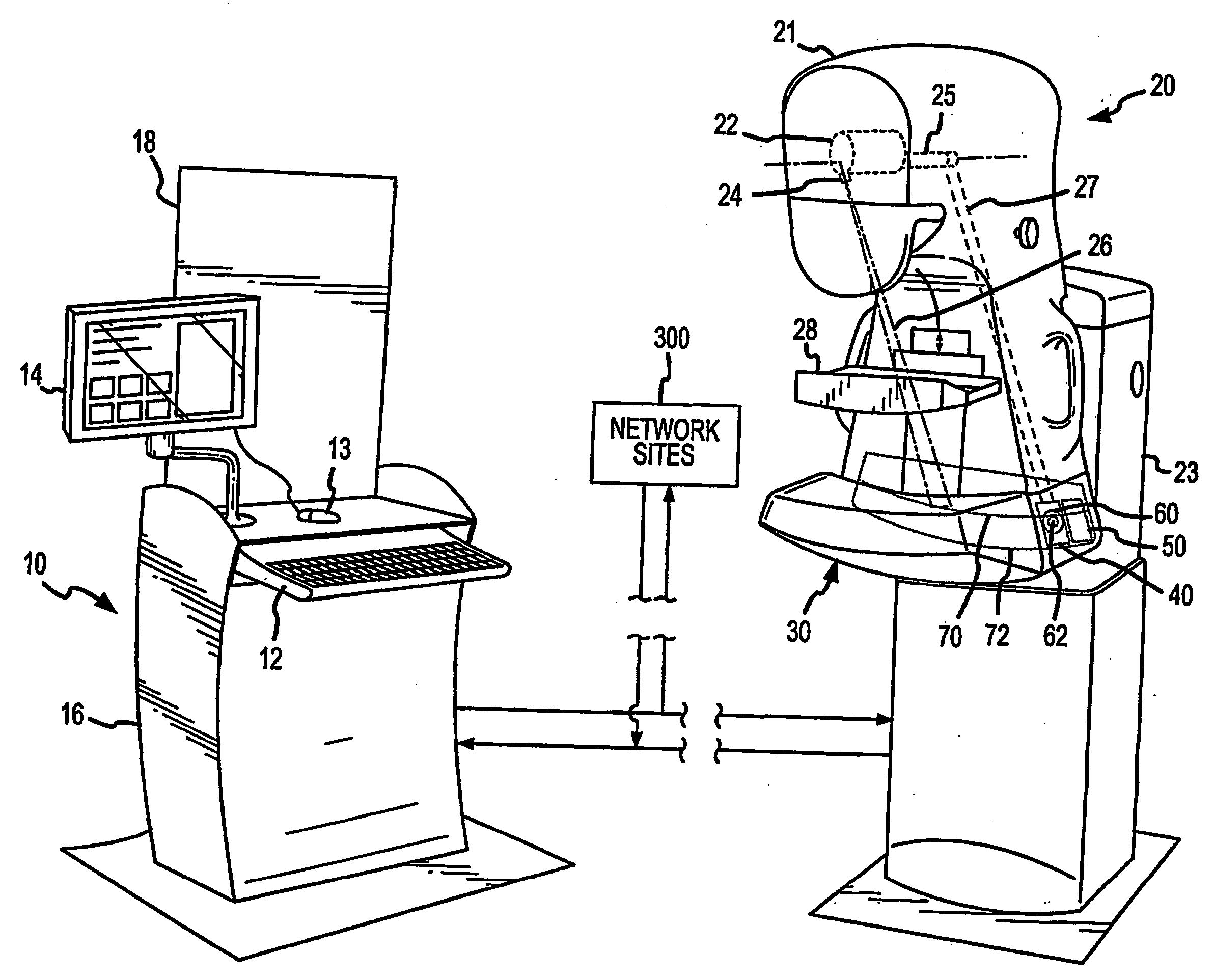
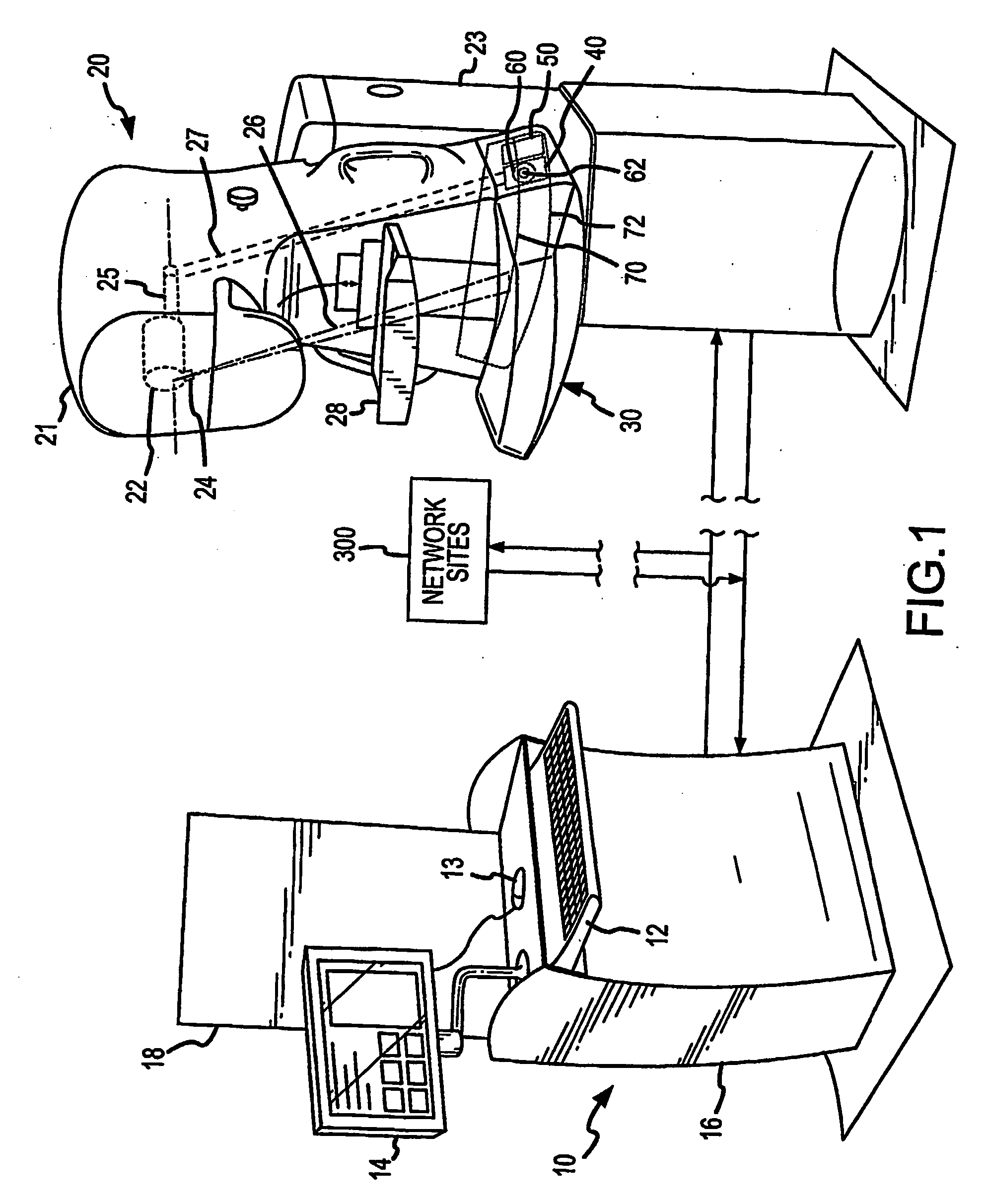
[0038]FIG. 1 illustrates one embodiment of an imaging system comprising the present invention. The system includes a monitoring station 10 and imaging station 20 operatively interconnected thereto, e.g. for patient screening and / or follow-up examination. The monitoring station 10 includes a user input keyboard 12 (e.g. for entering patient data), a display 14 and corresponding user input mouse 13 (e.g. for displaying / selecting images), and a processor 16 interconnected to the user input keyboard 12, display 14 and imaging station 20. Processor 16 is adapted to receive, process and store image data comprising image signals generated at the imaging station 20, and to control various operations at the imaging station 20. The monitoring station 10 may also include a radiopaque and optically transparent shield 18 for shielding medical personnel during observed patient imaging operations at the imaging station 20.

[0039] The monitoring station 10 and / or imaging station 20 may be further i...


PUM


Abstract

An integrated x-ray and ultrasound medical imaging system is provided, wherein a radiation detection means and ultrasound transducer may be disposed for scanning movement for image acquisition along either the same or substantially coincidental paths. The radiation detection means and ultrasound transducer may be advantageously located on the same side of the imaged body portion: The x-ray and ultrasound imaging operations may be sequential, partially overlapping, or synchronous. By virtue of the noted arrangement, increased accuracy and medical efficiencies can be realized.

Description

FIELD OF THE INVENTION [0001] The present invention relates to medical imaging systems, and more particularly, to an improved system that combinatively employs x-ray imaging and ultrasound imaging in a manner thai yields enhanced accuracy and multiple efficiencies. BACKGROUND OF THE INVENTION [0002] The advantages of early detection of potential lesions and suspicious masses within bodily tissue have been well-established. Increasingly, screening for common cancers of the breast, lung, colon, and prostate has gained support and acceptance in the medical community, but improvements in the sensitivity and specificity of the techniques remain key and are readily identifiable objectives. [0003] Of particular interest is the area of mammographic screening. After a given age or maturity, normally beginning at age 40, it is common for women to undergo periodic examinations, wherein film-based and / or digital x-ray screening mammograms are obtained. While significant advances have been made,...

Claims


Application Information

Patent Timeline
Patent Type & Authority Applications(United States)
IPC IPC(8): A61B6/03A61B6/00A61B8/08
CPCA61B6/488A61B6/502A61B6/5247A61B6/4417A61B8/0825A61B8/4416A61B8/565A61B6/563
Inventor BESSON, GUYNIELDS, MORGAN
Owner BESSON GUY
Why Eureka
Try Eureka