

Computer-aided image diagnostic processing device and computer-aided image diagnostic processing program product
a technology of image diagnostic and processing device, which is applied in the field of computer-aided image diagnostic processing device and computer-aided image diagnostic processing program product, can solve the problems of lung cancer examination effectiveness but small, multi-detector helical ct also has a problem of considerably increasing the burden of diagnosing reading, and nodule and lung blood vessel cannot be accurately distinguished from each other
- Summary
- Abstract
- Description
- Claims
- Application Information
AI Technical Summary
Problems solved by technology
Method used
Image

Examples
first embodiment
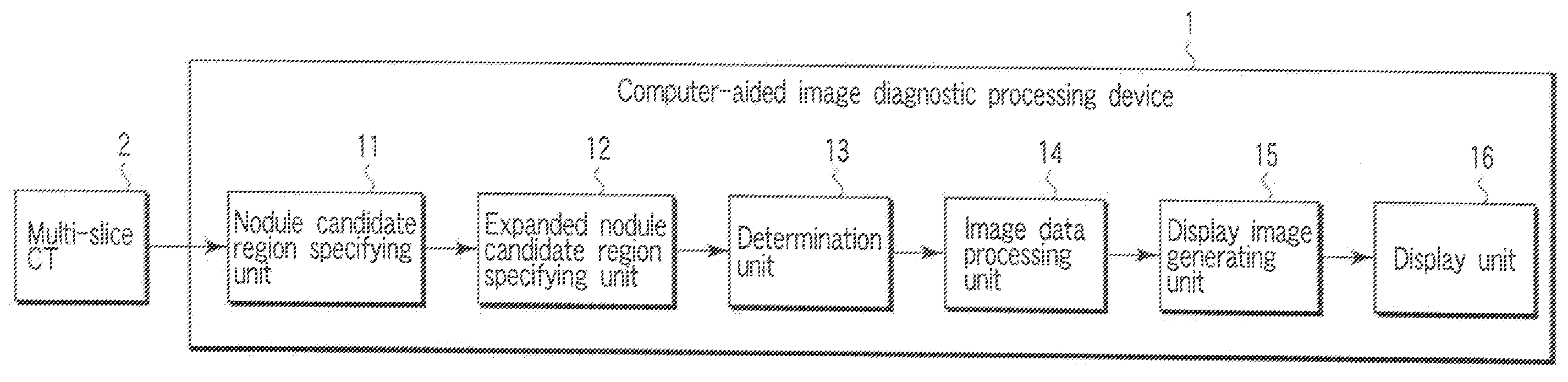
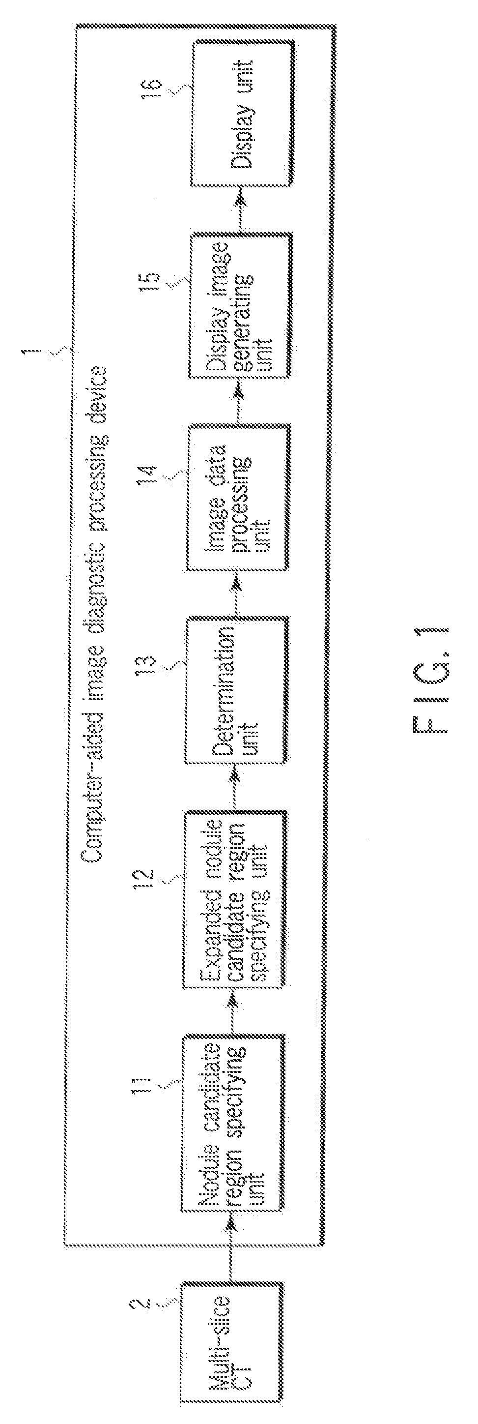
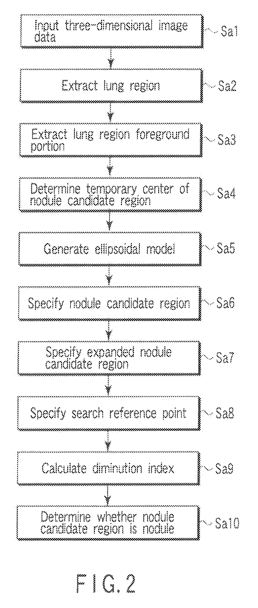
[0058]FIG. 1 is a view showing a structure of a computer-aided image diagnostic processing device 1 according to a first embodiment.
[0059]The computer-aided image diagnostic processing device 1 depicted in FIG. 1 is intended to process three-dimensional data acquired by a multi-slice CT 2. As shown in FIG. 1, the computer-aided image diagnostic processing device 1 includes a nodule candidate region specifying unit 11, a expanded nodule candidate region specifying unit 12, a determination unit 13, an image data processing unit 14, a display image generating unit 15, and a display unit 16.
[0060]The computer-aided image diagnostic processing device 1 can use, e.g., a general-purpose computer device as basic hardware. Further, the nodule candidate region specifying unit 11, the expanded nodule candidate region specifying unit 12, the determination unit 13, the image data processing unit 14, and the display image generating unit 15 can be realized when a processor mounted on the computer...
second embodiment
[0138]FIG. 23 is a view showing a structure of a computer-aided image diagnostic processing device 3 according to a second embodiment. It is to be noted that like reference numbers denote parts equal to those in FIG. 1, thereby omitting a detailed explanation thereof.
[0139]The computer-aided image diagnostic processing device 3 shown in FIG. 23 processes three-dimensional data acquired by a multi-slice CT 2. The computer-aided image diagnostic processing device 3 includes a nodule candidate region specifying unit 11, a expanded nodule candidate region specifying unit 12, a determination unit 13, a display unit 16, an input section 31, and a display image generating unit 32. That is, the computer-aided image diagnostic processing device 3 includes the display image generating unit 32 in place of the display image generating unit 15 in the computer-aided image diagnostic processing device 1, and further includes the input section 31. The computer-aided image diagnostic processing devi...
third embodiment
[0160]Since a structure of a computer-aided image diagnostic processing device according to a third embodiment is the same as that of the computer-aided image diagnostic processing device 3 according to the second embodiment, thereby omitting an illustration thereof. Further, FIG. 23 is used for an explanation of the third embodiment.
[0161]The third embodiment is different from the second embodiment in processing carried out by a display image generating unit 32. That is, the display image generating unit 32 according to the third embodiment is realized by changing a medical diagnostic imaging assistance program executed by a processor mounted on a computer device used as basic hardware.
[0162]Operation of the computer-aided image diagnostic processing device 3 according to the third embodiment will now be explained.
[0163]A nodule candidate region and its peripheral region are specified by a nodule candidate region specifying unit 11, a expanded nodule candidate region specifying reg...
PUM
 Abstract
Description
Claims
Application Information