Ultrasound medical imaging with robotic assistance for volume imaging

Inactive Publication Date: 2008-01-24
SIEMENS MEDICAL SOLUTIONS USA INC
View PDF50 Cites 104 Cited by
  • Summary
  • Abstract
  • Description
  • Claims
  • Application Information

AI Technical Summary

Benefits of technology

[0004]By way of introduction, the preferred embodiments described below include methods, systems and computer readable media for ultrasound imaging with robotic assistance. A robotic mechanism positions a volume scanning transducer at multiple acoustic windows on a patient. Ultraso

Problems solved by technology

Since the transducer has only a limited field of view, the sonographer spends a lot of time trying to find the area of interest.
Scanning by a sonographer is expensive and prone to variability and human e

Method used


Image

  • Ultrasound medical imaging with robotic assistance for volume imaging
  • Ultrasound medical imaging with robotic assistance for volume imaging
  • Ultrasound medical imaging with robotic assistance for volume imaging

Examples


Example

DETAILED DESCRIPTION OF THE DRAWINGS AND PRESENTLY PREFERRED EMBODIMENTS

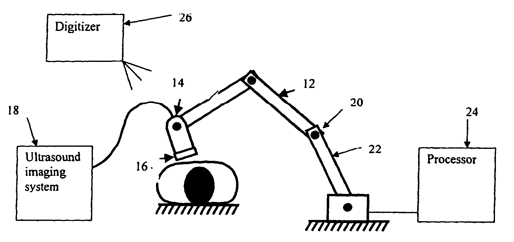
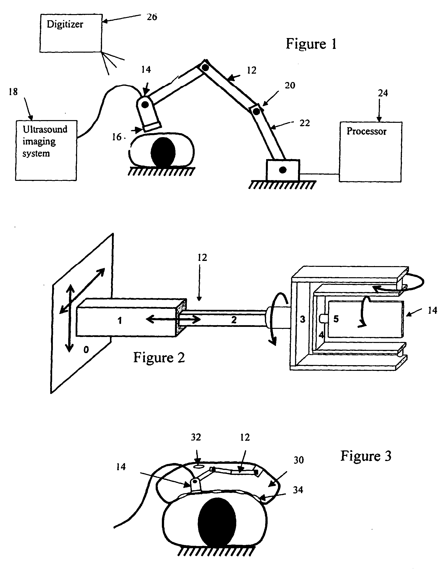
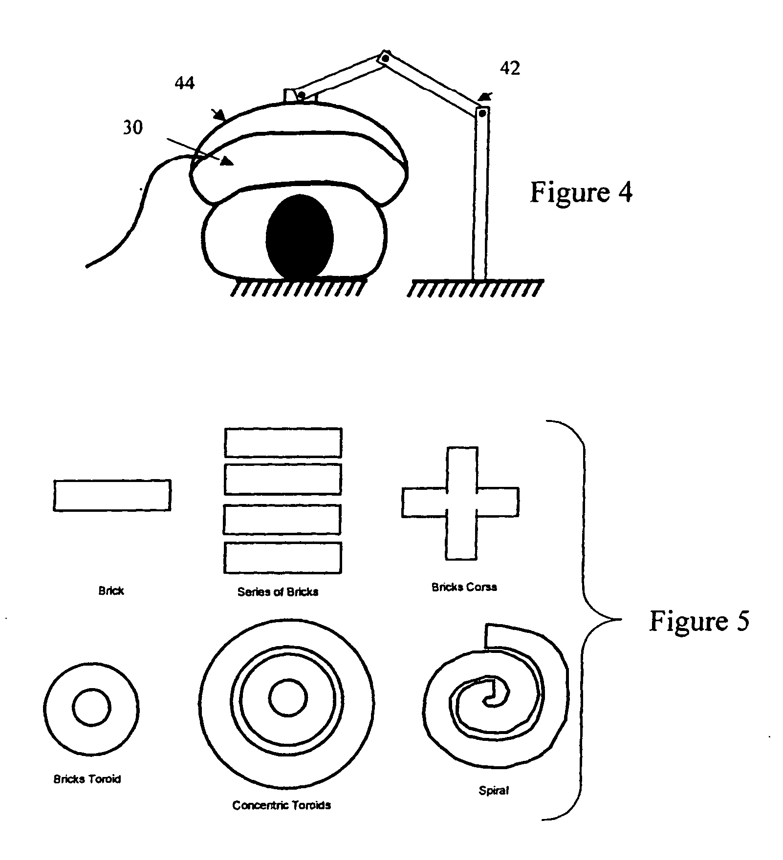
[0020]A robotic mechanism assists with ultrasound imaging. The robotic mechanism connects with a volume transducer, such as a wobbler or multi-dimensional array. In one form of assistance, the robotic mechanism repositions the transducer to a plurality of locations for a wide-region or body type (full, torso or abdomen) scan. The volume data from each position is combined into a data set for analysis. In another form of assistance, the sonographer controls placement of the transducer by hand, but the robotic mechanism applies force in a direction indicated by the user. The user may have less strain due to the assistance by the robotic mechanism.

[0021]One or more ultrasonic probes connect with one or more robotic manipulator arms, force sensors and position sensors. A robotic manipulator arm is a mechanical device containing a series of links connected by active joints. Each joint may have a motor and a sensor se...


PUM


Abstract

A robotic mechanism positions a volume scanning transducer at multiple acoustic windows on a patient. Ultrasound data is acquired from the windows and combined into a wide field-of-view. The robotic mechanism operates without user contact, such as for an automated full or partial torso scan of a patient. Alternatively, the robotic mechanism provides force to reduce strain on a sonographer.

Description

BACKGROUND[0001]The present embodiments relate to ultrasound imaging. In particular, a robot assists with ultrasound imaging.[0002]A sonographer holds a transducer for ultrasound imaging. Holding the transducer has several drawbacks. Since the transducer has only a limited field of view, the sonographer spends a lot of time trying to find the area of interest. Once the area of interest is found, only the area of interest is scanned. If there is an additional area to be investigated, the patient typically has a separate appointment for additional scanning. In contrast, computed tomography and magnetic resonance imaging use a gantry to slide the patient in and out of the scanner, acquiring data from a large area. All the data needed for the physician to diagnose the disease may be acquired during a single session.[0003]Image quality of ultrasound depends on the sonographer and how much pressure is applied by the sonographer to the transducer against the patient. Scanning by a sonograp...

Claims


Application Information

Patent Timeline
IPC IPC(8): A61B8/00
CPCA61B8/4209A61B8/483A61B8/4281A61B8/4218
Inventor SUMANAWEERA, THILAKA
Owner SIEMENS MEDICAL SOLUTIONS USA INC
Why Eureka
Try Eureka