
System and method for coronary segmentation and visualization

a multi-branched, coronary vein technology, applied in the field of visualizing and segmenting multi-branched coronary veins, can solve the problems of difficult to achieve accurate segmentation of coronary veins, inconvenient use of these approaches, and inability to accurately segment the coronary vein, etc., to achieve the effect of eliminating incorrect edges

Inactive Publication Date: 2008-05-01
SIEMENS HEALTHCARE GMBH
View PDF13 Cites 69 Cited by
  • Summary
  • Abstract
  • Description
  • Claims
  • Application Information

AI Technical Summary

Benefits of technology

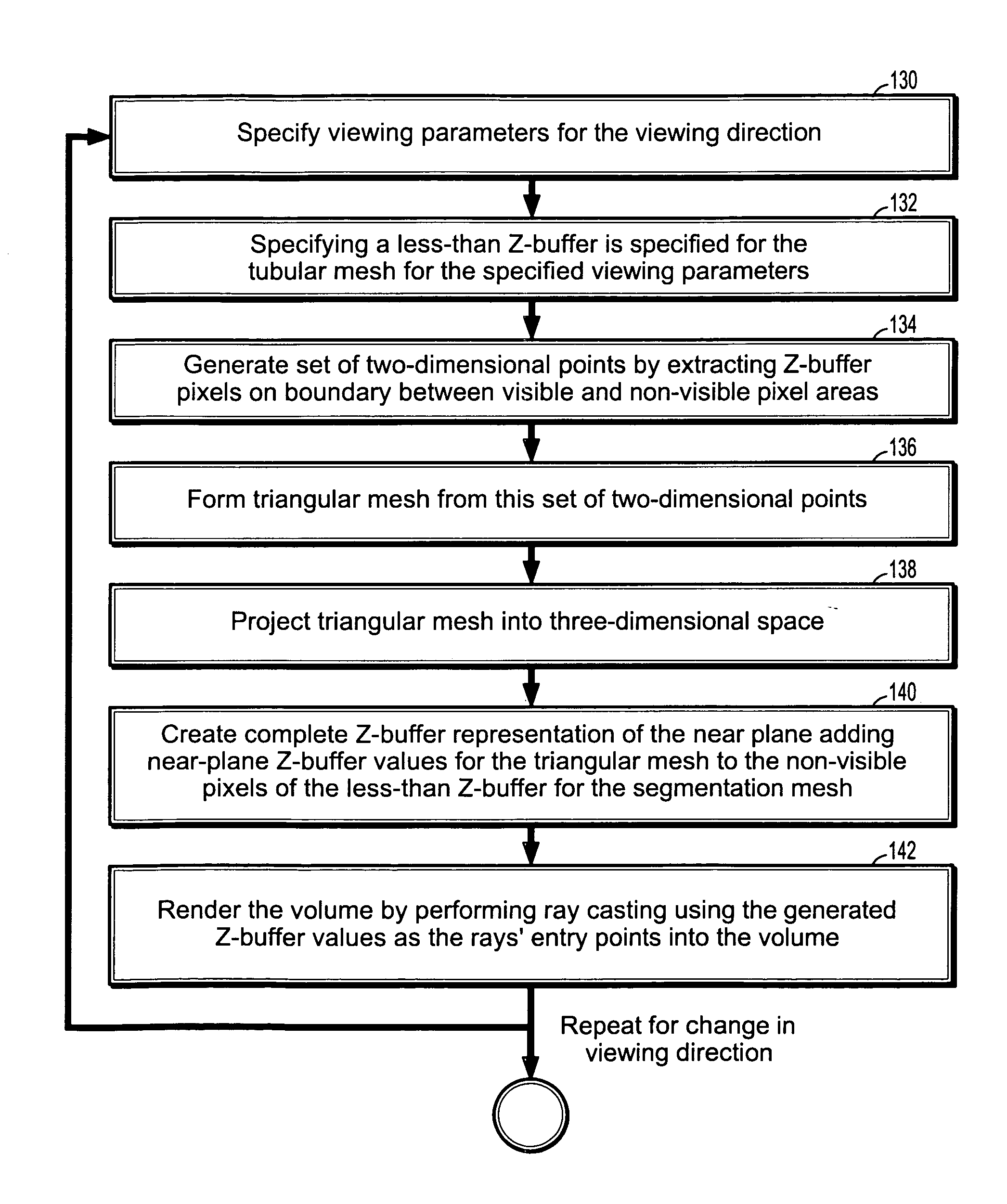
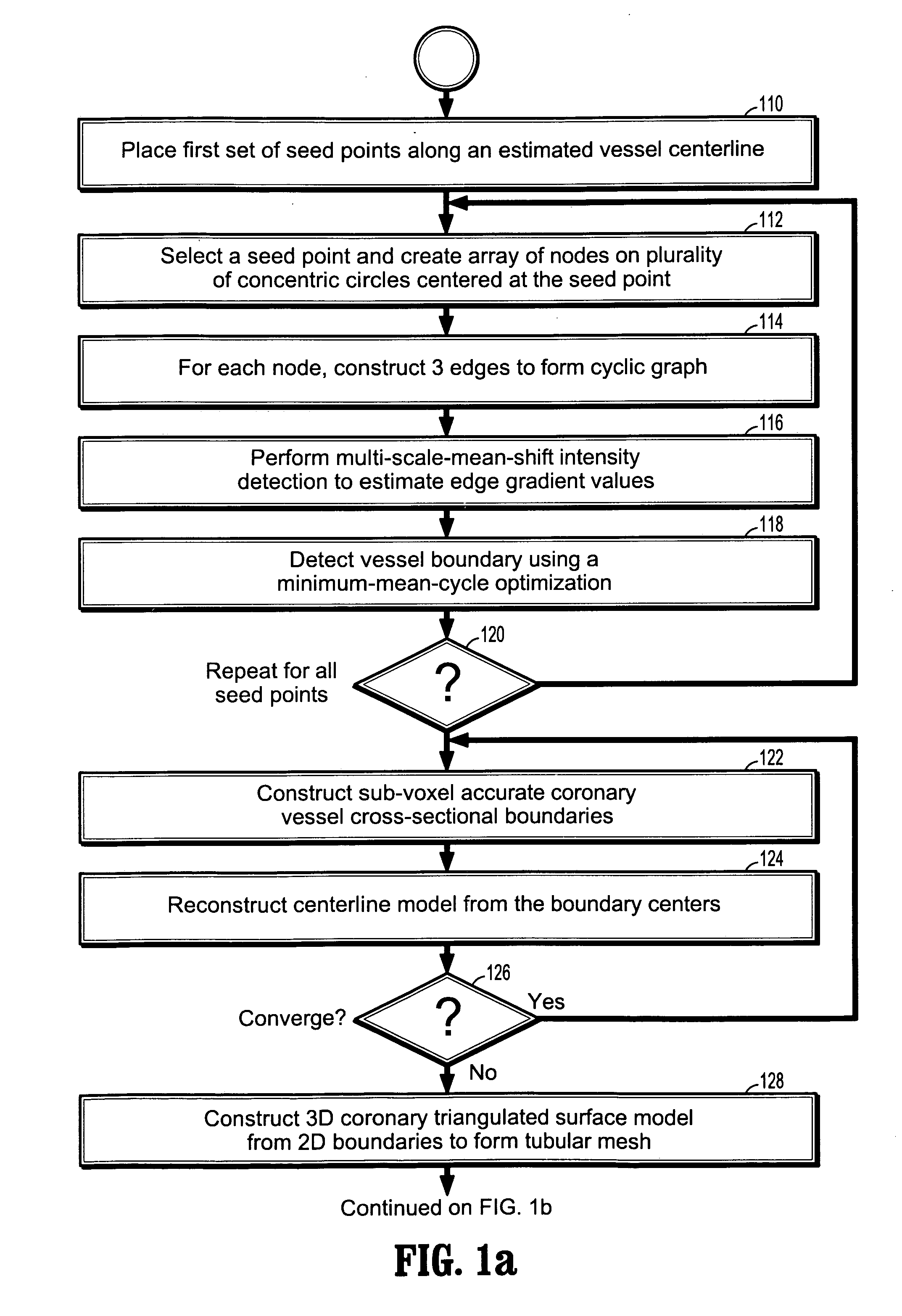
[0013]According to a further aspect of the invention, performing a multi-scale-mean shift filtering comprises propagating one or more rays along the vessel, representing each ray as a collection of 2-dimensional points, performing a mean-shift filtering to each 2D points until convergence, selecting a plurality of edges based on a confidence function of the mean-shift filter size, and eliminating incorrect edges.

Problems solved by technology

However, most of these approaches are not suited for coronary vessels, especially when the seed point falls near or outside of a vessel boundary.
Accurate segmentation of coronary arteries is challenging since the physical size of the coronary vessels is quite small and is sensitive to noise and partial volume effects.
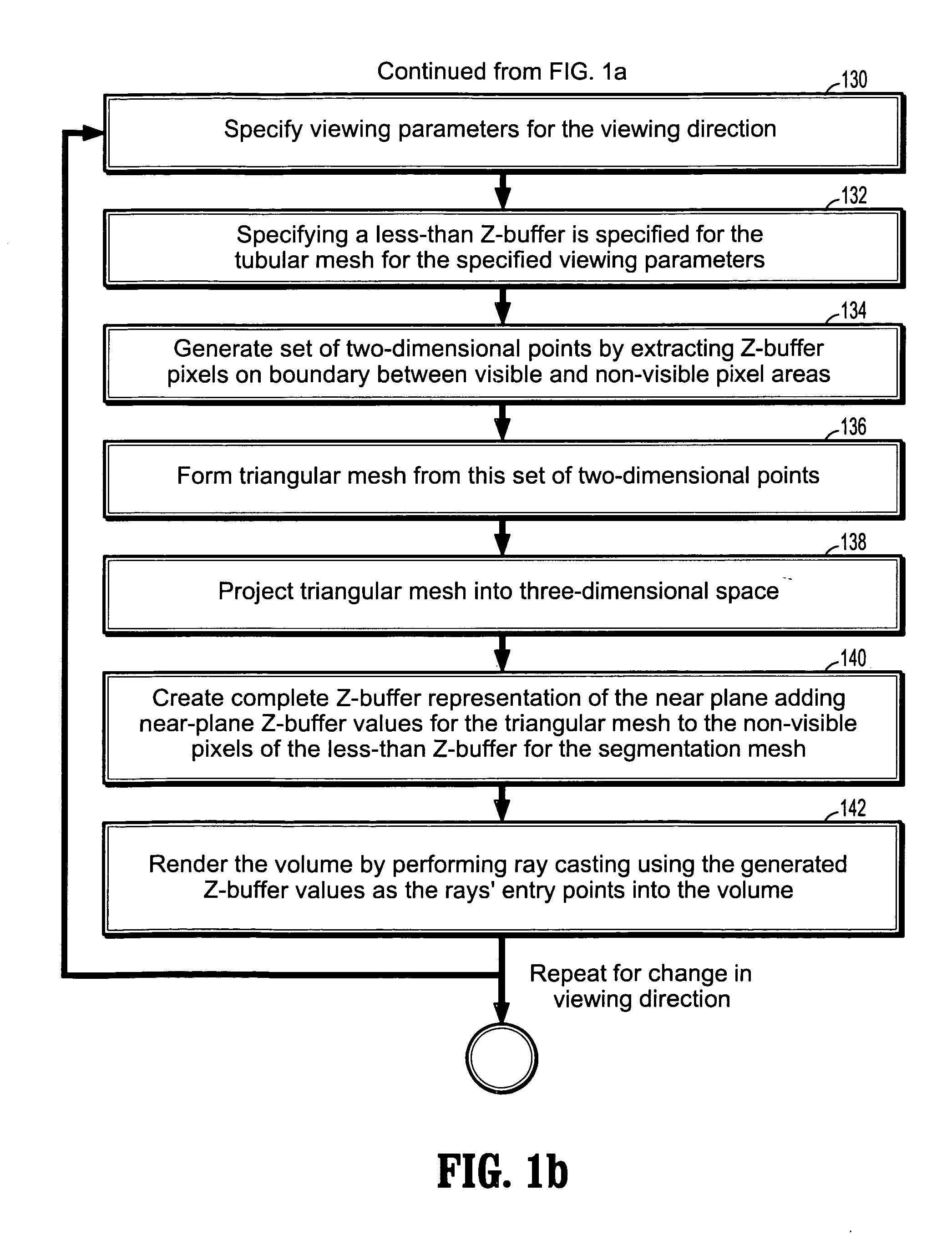
Effective visualization of the segmented coronaries is also challenging to achieve.
For example, a direct three-dimensional (3D) surface rendering may involve the risk of obliterating stenoses.
Furthermore, this rendering technique completely masks out of the image the area surrounding these vessels.
This lack of surrounding area information significantly reduces the effectiveness of the visualization, and obscures the exact location and orientation of the visualized vessels.
Accurate and robust detection of vessel boundaries is another challenging task.
If the vessel boundary detection algorithm is inaccurate, even in a small number of cases, a medical professional (e.g., a radiologist) relying on the computer's output may, in turn, incorrectly diagnose the patient.
There are many reasons why accurate and robust detection of vessel boundaries is a challenging task.
First, the presence of significant noise levels in computed tomography (CT) and magnetic resonance (MR) images often forms strong edges, i.e. changes in intensity between data points, inside vessels.
Second, the size of a vessel can vary from one vessel location to another, resulting in additional edges.
Fourth, the presence of vascular pathologies, such as calcified plaques, often makes the shape of a vessel cross-sectional boundary locally deviate from a circular shape.

Method used


Image

  • System and method for coronary segmentation and visualization
  • System and method for coronary segmentation and visualization
  • System and method for coronary segmentation and visualization

Examples


Embodiment Construction

[0030]Exemplary embodiments of the invention as described herein generally include systems and methods for visualization and segmentation of multi-branched coronary vessels. Accordingly, while the invention is susceptible to various modifications and alternative forms, specific embodiments thereof are shown by way of example in the drawings and will herein be described in detail. It should be understood, however, that there is no intent to limit the invention to the particular forms disclosed, but on the contrary, the invention is to cover all modifications, equivalents, and alternatives falling within the spirit and scope of the invention.

[0031]As used herein, the term “image” refers to multi-dimensional data composed of discrete image elements (e.g., pixels for 2-D images and voxels for 3-D images). The image may be, for example, a medical image of a subject collected by computer tomography, magnetic resonance imaging, ultrasound, or any other medical imaging system known to one o...


PUM


Abstract

A method of coronary vessel segmentation and visualization includes providing a digitized coronary image, placing a plurality of seed points along an estimated centerline of a coronary vessel, selecting a seed point and constructing a cyclic graph around the seed point in a plane perpendicular to the centerline at the seed point, performing a multi-scale-mean shift filtering in the perpendicular plane to estimate image gradient values, detecting a vessel boundary using a minimum-mean-cycle optimization that minimizes a ratio of a cost of a cycle to a length of a cycle, constructing a sub-voxel accurate vessel boundary about a point on the centerline, and refining the location of the centerline point from the sub-voxel accurate boundary, where the steps of constructing a sub-voxel accurate vessel boundary and refining the centerline point location are repeated until convergence.

Description

CROSS REFERENCE TO RELATED UNITED STATES APPLICATIONS[0001]This application claims priority from “Advanced Coronary Visualization and Segmentation”, U.S. Provisional Application No. 60 / 854,183 of Aharon, et al., filed Oct. 26, 2006, the contents of which are herein incorporated by reference.TECHNICAL FIELD[0002]This disclosure is directed to the visualization and segmentation of multi-branched coronary vessels.DISCUSSION OF THE RELATED ART[0003]Non-invasive diagnosis of coronary artery pathologies has become a reality due to the technological advances in image acquisition devices such as the introduction of multi-detector computed tomography (CT). While these advances have significantly improved image quality, the diagnosis of such pathologies still requires advanced segmentation, quantification and visualization algorithms. There are many cross-sectional vessel segmentation algorithms known in the art. However, most of these approaches are not suited for coronary vessels, especiall...

Claims


Application Information

Patent Timeline
IPC IPC(8): G06T17/00
CPCG06T7/0083G06T7/0093G06T7/60G06T2207/30101G06T2207/10072G06T2207/20044G06T17/20G06T7/12G06T7/162
Inventor AHARON, SHMUELGULSUN, MEHMET AKIFTEK, HUSEYIN
Owner SIEMENS HEALTHCARE GMBH
Learn More