Radiological imagery method making a zone of interest in an organ correspond to an associated part of the network

Active Publication Date: 2008-05-29
GENERAL ELECTRIC CO
View PDF3 Cites 8 Cited by
  • Summary
  • Abstract
  • Description
  • Claims
  • Application Information

AI Technical Summary

Benefits of technology

[0018]This method allows the operator to identify the part of the network that participates in supplying or leaving the zone of interest, and only this zone.
[0019]Th

Problems solved by technology

In this type of operation, the radiologist must access an operation point that will give an optimum result, in other words treatment of the entire target zone and also limitation of the impact of the operation on the surrounding tissues.
This task is usually

Method used


Image

  • Radiological imagery method making a zone of interest in an organ correspond to an associated part of the network
  • Radiological imagery method making a zone of interest in an organ correspond to an associated part of the network
  • Radiological imagery method making a zone of interest in an organ correspond to an associated part of the network

Examples


Example

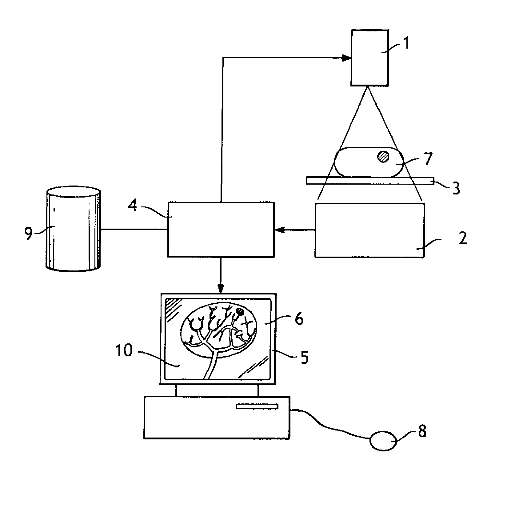
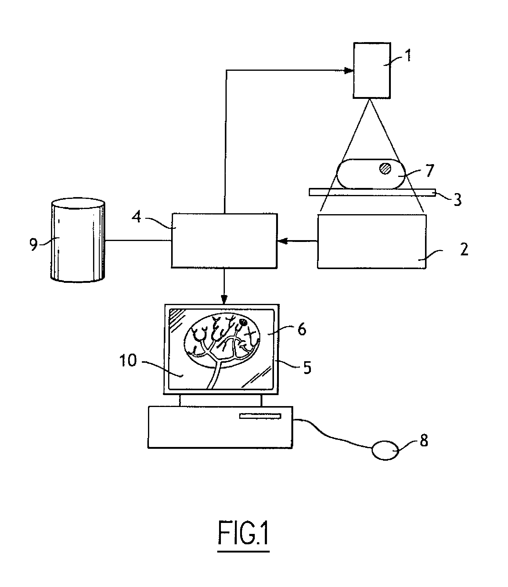
[0049]The imagery apparatus shown in FIG. 1 comprises a source 1 capable of emitting an X-ray beam 8, a detector 2 placed facing the source 1 and capable of detecting rays emitted by source 1, a support 3 placed between the source 1 and the detector 2.

[0050]The support 3 may receive an organ 7 for which an image is to be acquired.

[0051]The apparatus comprises a processing unit 4 (for example a computer) capable of receiving data supplied by the detector 2 and that can control the source 1 and the detector 2. The treatment unit 4 can control the emission of X-rays by the source and reading of an image by the detector 2.

[0052]The apparatus comprises an interface unit 5 comprising a screen 6 and control means including a mouse 8.

[0053]Finally, the apparatus comprises a database 9 in which images of the organ 7 that were previously acquired are saved.

[0054]The processing unit 4 can control the interface unit so that the interface unit displays an image of the organ 7 acquired in real ti...


PUM


Abstract

Embodiments of the invention relate to a method of processing a radiological image of an organ, the organ being connected to a flow circulation network, and comprising steps according to which:
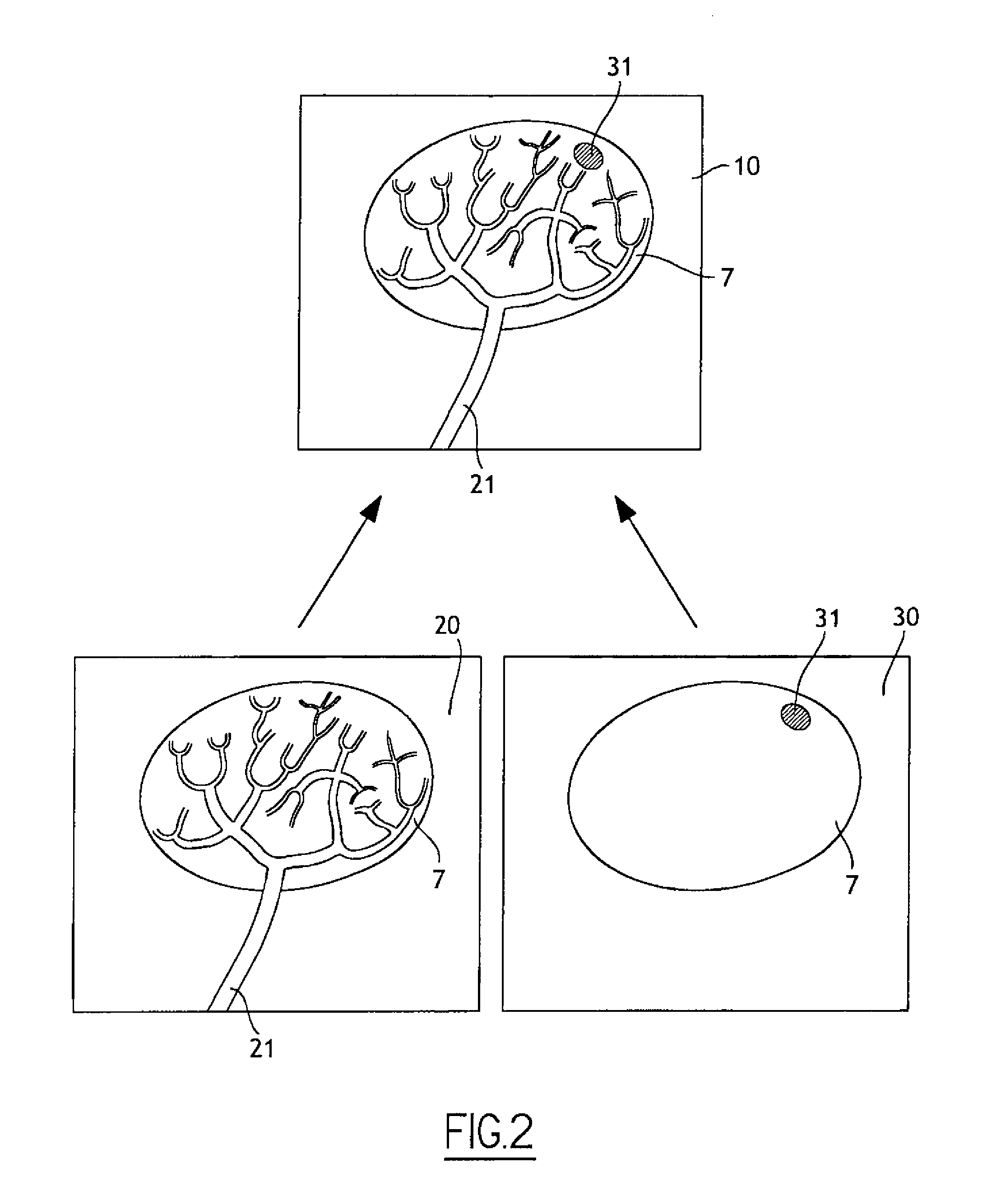
    • an operator or a processing means defines a contour to delimit a zone of interest in the radiological image of the organ;
    • the processing means determines part of the network in which the flow supplies the zone of interest or originates from this zone of interest; and
    • a display means displays the determined part of the network.

Description

FIELD OF THE INVENTION[0001]The field of the invention relates to the domain of radiological imagery, and more particularly the domain of operational radiological imagery.STATE OF THE ART[0002]Operational radiology denotes surgical operation techniques in which a radiologist displays images of an organ provided by an imagery apparatus to guide or control an operation on this organ.[0003]These techniques are particularly useful for performing surgical operations that follow natural circulation pathways in the body (for example the blood, bile and lymph networks) to access a zone to be treated. The surgeon introduces a catheter into a vein or artery until reaching an appropriate point, using an image output by the imagery apparatus.[0004]These techniques have the advantage that they are not very invasive compared with traditional surgical techniques.[0005]For example, operational radiology can be used to treat a target tissue zone by injecting a substance or by closing off a vessel at...

Claims


Application Information

Patent Timeline
IPC IPC(8): A61B6/00G06K9/62
CPCA61B6/507G06T7/0012
Inventor PICHON, ERICTROUSSET, YVESKNOPLIOCH, JEROME
Owner GENERAL ELECTRIC CO
Why Eureka
Try Eureka