Freeze-dried platelets as a diagnostic agent

a platelet and freeze-dried technology, applied in the field of medicine, can solve the problems of excessive blood loss, clot formation may be compromised, occluded, etc., and achieve the effect of fast response, superior response to agonists, and rapid respons

Active Publication Date: 2008-06-19
CELLPHIRE INC
View PDF6 Cites 19 Cited by
  • Summary
  • Abstract
  • Description
  • Claims
  • Application Information

AI Technical Summary

Benefits of technology

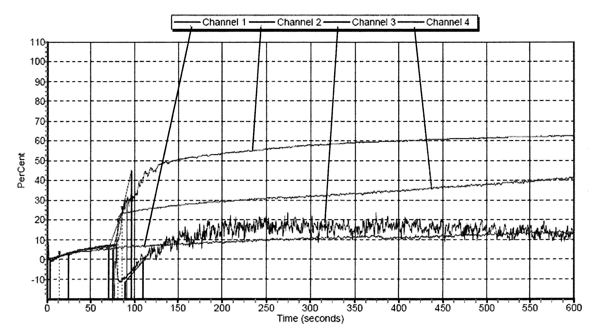
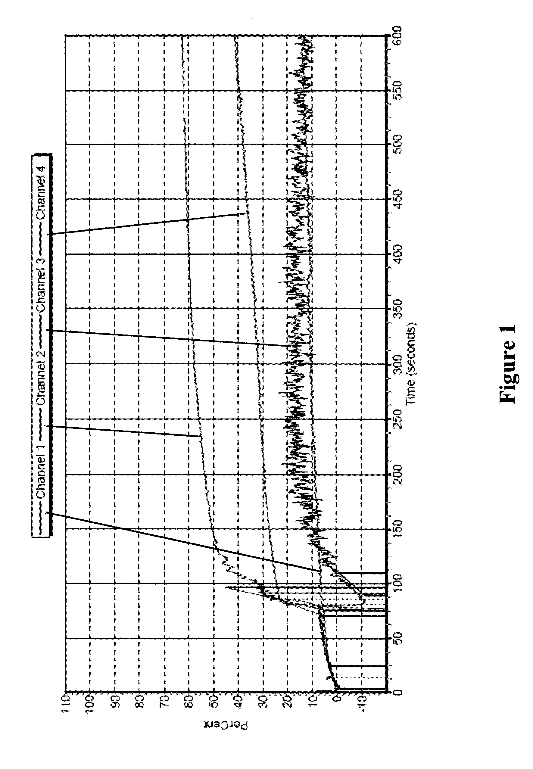
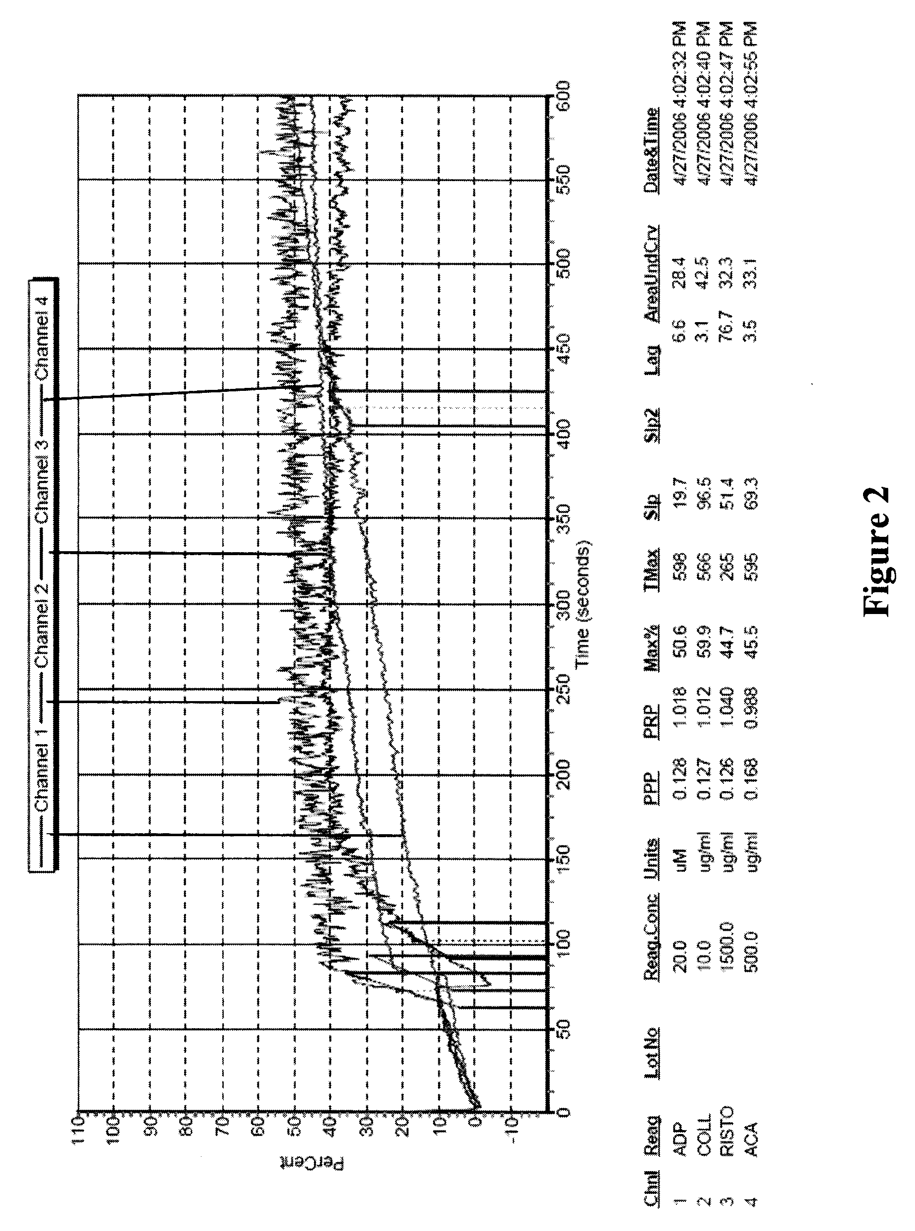
[0018]In one aspect, the invention provides freeze-dried platelets. In general, the freeze-dried platelets of the present invention have all or essentially all of the surface markers that are known to be relevant to blood clotting and that are present on platelets found in vivo or on freshly obtained platelets. In contrast to other freeze-dried platelets known in the art, the freeze-dried platelets of the present invention have the ability to rapidly respond to normal platelet agonists. For example, they can rapidly respond to agonists currently being used to assess platelet activation functions, such as thrombin, thrombin activation peptide (TRAP), arachidonic acid, collagen, ADP, ristocitin, and epinephrine. Thus, they can be considered to have superior responses to agonists, as compared to freeze-dried platelets known in the art. In addition, as compared to freeze-dried platelets known in the art, the freeze-dried platelets of the present invention quickly produce, upon exposure to one or more agonists, aggregates of a size sufficient to detect using standard light transmission aggregometry. Like the compositions discussed below, the platelets of the invention can be used as a control reagent for test methods that are currently in use for testing the activity of blood and blood components. They also can be used as a control reagent for calibrating instruments, such as light transmission aggregometers.

Problems solved by technology

Blood clotting is a complicated process: if the clot formation is unchecked, the vessel will become occluded; if the clot is not sturdy, excessive blood loss will occur.
In situations where normal hemostasis is unbalanced, clot formation may be compromised.
Moreover, these assays are potentially unreliable as they are end-point tests in which results are based on the time of clot formation in vitro.
Another limitation relates to the fact that exogenous reagents, such as kaolin, thrombin, calcium, etc. must be added; thus, the results are based on an artificial system, and do not necessarily reflect the patient's thrombotic potential.
Dysfunctional platelets can lead to abnormal hemorrhage, such as bleeding or thrombosis.
However, such analyses, although accurate, are not highly sensitive, and can fail to detect slight perturbations in normal clotting function at early stages of a disorder.

Method used


Image

  • Freeze-dried platelets as a diagnostic agent
  • Freeze-dried platelets as a diagnostic agent
  • Freeze-dried platelets as a diagnostic agent

Examples


example 1

Initial Preparation of Reagents and Platelets

[0116]Buffers and Solutions:

[0117]Suspension Buffer:

[0118]9.5 mM HEPES

[0119]75 mM NaCl

[0120]4.8 mM KCl

[0121]5 mM glucose

[0122]12 mM NAHCO3

[0123]100 mM Trehalose

[0124]1% EtOH

[0125]Stock Solutions and Reagents:

[0126]30% Polysucrose Solution (12 g Polysucrose Ficoll™ PM400 in 40 ml water)

[0127]1×PBS (standard solution)

[0128]PBSE, pH 7.4 (PBS plus 10 mM EDTA)

[0129]PBSE, pH 6.5

[0130]Remove all buffers and other reagents from refrigerators, freezers, and other storage areas. Allow each to reach ambient temperature (approximately 21° C.-25° C.). All buffers that require daily preparation should be made while previously prepared buffers are warming. When platelet units arrived, they should be placed on an orbital shaker reciprocating at approximately 50 RPM until use.

example 2

Initial Preparation of Pooled Plasma

[0131]If using more than one unit of plasma, pool all acceptable units of platelet rich plasma (PRP) in one large plastic beaker. Take a platelet cell count of the undiluted pooled PRP on an instrument suitable for platelet counting, such as the AC·T 10 Coulter counter. Stir and measure the pH of the PRP. If the pH is greater than 6.8, adjust the pH to between 6.4 and 6.8 with ACD. If the pH adjustment is overshot (too much ACD added) to between 6.2-6.4, verify that the platelets are still swirling and if so, continue to the next step.

[0132]If the original platelet source was an apheresis unit, skip the following step. If the original platelet source was a random donor unit (RDU), perform the following RBC removal step. Divide PRP equally between 50 ml conical tubes. Spin the PRP in a swinging bucket centrifuge for 3 to 5 minutes at 750×g at room temperature. Remove the PRP and pool in a clean plastic beaker, leaving behind the pellet of RBCs at t...

example 3

Preparation of Lyophilized Platelets

[0136]DMSO Incubation

[0137]Resuspend platelets gently in a small volume of PBSE, pH7.4, taking care not to mix vigorously, resulting in activation of platelets. Where possible, use gentle “puffs” of PBSE, pH 7.4 to resuspend the platelets.

[0138]Add 10 ul of DMSO for every 1×10⁹ platelets total.

[0139]Incubate platelets with DMSO at room temperature for 30 minutes while shaking gently on an orbital shaker (˜50 RPM).

[0140]After incubation, gently add a large volume of PBSE, pH6.5 to the platelet samples with a transfer pipette until the samples reach a pH of 6.5-6.7.

[0141]Aliquot out the resuspended platelets evenly between clean conical centrifuge tubes.

[0142]Centrifuge the platelets in a swinging bucket centrifuge at 1000×g for 10 minutes at room temperature. After the spin, remove the supernatant, leaving the platelet pellet in the bottom of the conical tube.

[0143]Platelet Loading

[0144]Resuspend the platelet pellet in a minimal volume of Suspensio...


PUM


Abstract

The present invention provides freeze-dried platelets and compositions comprising freeze-dried platelets for use as a diagnostic and calibration reagent for blood coagulation function. The invention also provides methods of diagnosing or monitoring blood coagulation function, including diagnosing or monitoring blood coagulation diseases and disorders. Kits for performing the methods of the invention are also included.

Description

CROSS-REFERENCE TO RELATED APPLICATIONS[0001]This application relies on the disclosure of and claims the benefit of the filing date of U.S. provisional patent application No. 60 / 869,915, filed on 14 Dec. 2006, the entire disclosure of which is hereby incorporated herein by reference.BACKGROUND OF THE INVENTION[0002]1. Field of the Invention[0003]The present invention relates to the field of medicine. More specifically, it relates to blood and blood products, and uses of those products to identify diseases and disorders affecting the blood or components of it. In many instances, the present invention relates to the field of dry platelet preparations and their use in diagnosis and monitoring of diseases and disorders relating to platelet function.[0004]2. Description of Related Art[0005]Platelets are formed in the bone marrow as fragments of megakaryocytes. They are irregularly-shaped, colorless bodies that are present in blood at a concentration of 150,000-450,000 per microliter (ul)...

Claims


Application Information

Patent Timeline
Patent Type & Authority Applications(United States)
IPC IPC(8): A01N1/02C12N5/06C12N5/078
CPCA01N1/02C12N2503/00C12N5/0644A01N1/0221
Inventor HO, DAVIDJONES, HEATHER
Owner CELLPHIRE INC
Why Eureka
Try Eureka