Methods for performing a medical procedure

a medical procedure and method technology, applied in the field of medical procedures, can solve the problems of excessive intravenous injection, high risk of excess intravenous injection, and insufficient space in the uterus that is not naturally available in the uterus

Active Publication Date: 2008-06-19
HOLOGIC INC
View PDF102 Cites 82 Cited by
  • Summary
  • Abstract
  • Description
  • Claims
  • Application Information

AI Technical Summary

Problems solved by technology

Unfortunately, however, adequate space typically does not exist naturally in the uterus because the uterus is a flaccid organ.
For example, because the distending fluid is administered under pressure (which pressure may be as great as 120 mm Hg or greater), there is a risk that such fluids may be taken up by a blood vessel in the uterus, i.e., intravasation, which uptake may be quite harmful to the patient.
Because the risk of excess intravasation can lead to death, it is customary to monitor the fluid uptake on a continuous basis using a scale system.
This risk of excess intravasation is particularly great when the fluid distension technique is followed by a procedure in which a blood...

Method used


Image

  • Methods for performing a medical procedure
  • Methods for performing a medical procedure
  • Methods for performing a medical procedure

Examples


Embodiment Construction

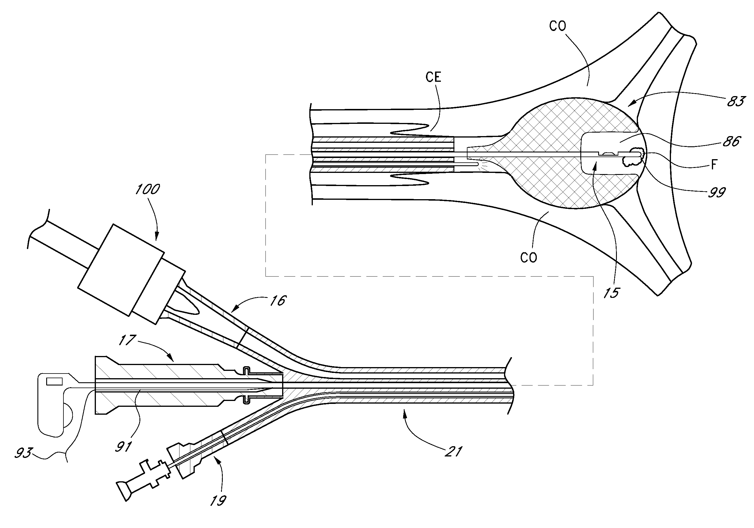
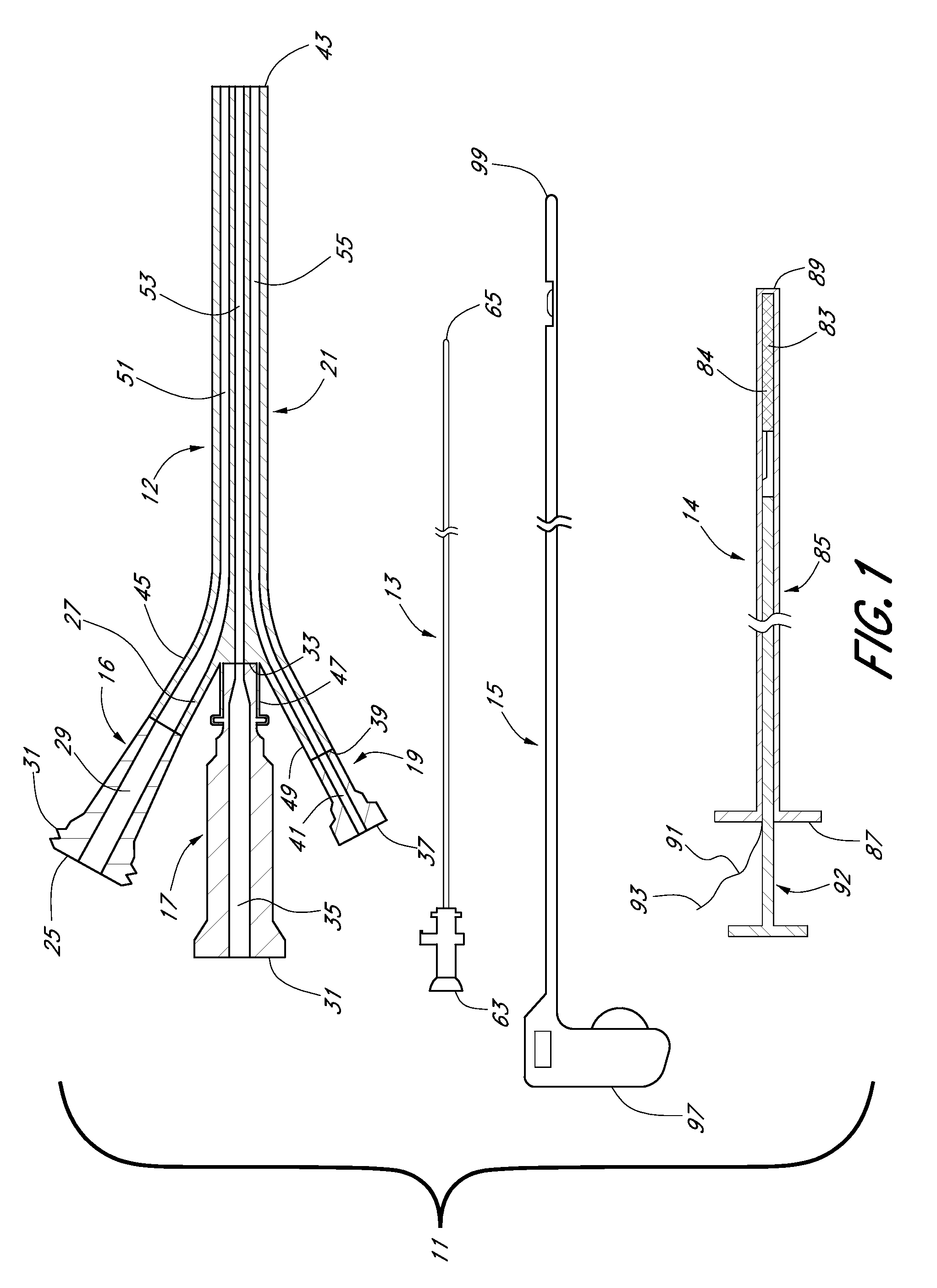
[0029]Referring now to FIG. 1, there is shown a plan view, partly in section, of one embodiment of a system that may be used in accessing and in examining and / or treating a body cavity, the system being constructed according to the teachings of the present invention and being represented generally by reference numeral 11.

[0030]System 11, which is shown in a partially disassembled state, is particularly well-suited for use in accessing and examining and / or treating the uterus of a female patient. However, it should be understood that system 11 is not limited to such a use and may be used in other anatomies that may be apparent to those of ordinary skill in the art.

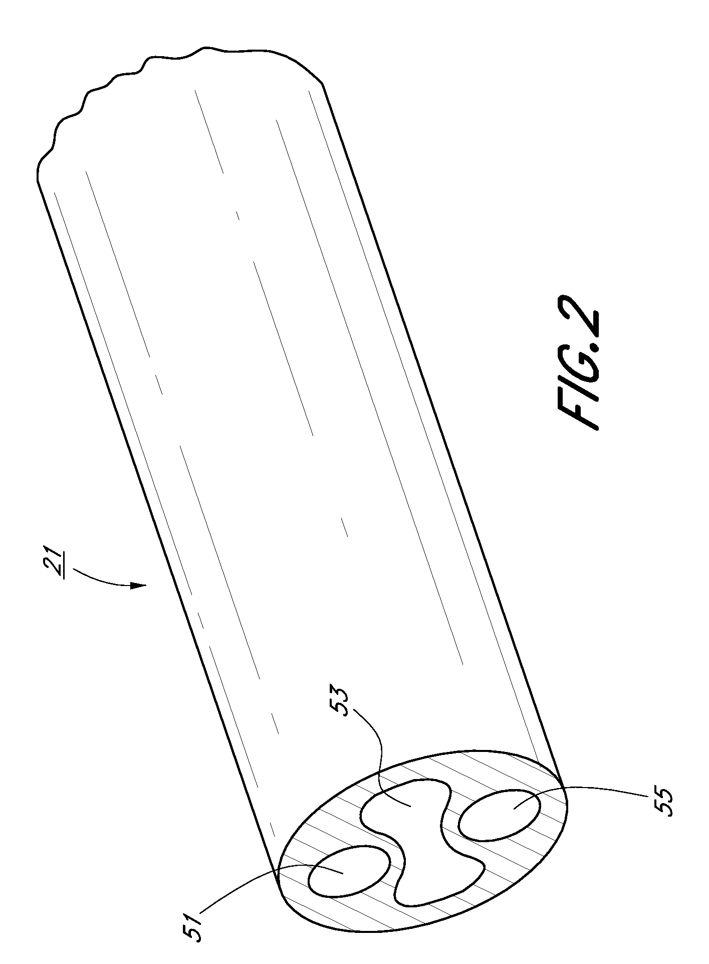
[0031]System 11 may comprise an introducer 12, a visualization device 13, a distension device 14 and a tissue modifying device 15. Introducer 12, in turn, may include a first port 16, a second port 17, a third port 19, and a flexible sheath 21. Ports 16, 17 and 19 are typically not intended for insertion into a patient wher...


PUM


Abstract

Methods are disclosed, for performing therapeutic or diagnostic procedures at a remote site. According to one embodiment, the methods include the use of a system including an introducer designed for transcervical insertion into the uterus. The introducer is constructed to include a fluid lumen, an instrument lumen, and a visualization lumen. The system may include a fluid source, which is coupled to the fluid lumen and is used to deliver a fluid to the uterus either for washing the uterus or for fluid distension of the uterus. The system additionally includes a tissue modifying device, such as a morcellator, and a distension device for distending the uterus and/or for maintaining the uterus in a distended state. The tissue modifying device and the distension device are alternately deliverable to the uterus through the instrument lumen. The system may further include a hysteroscope deliverable to the uterus through the visualization lumen.

Description

CROSS-REFERENCE TO RELATED APPLICATIONS[0001]The present application claims the benefit under 35 U.S.C. 119(e) of U.S. Provisional Patent Application Ser. No. 60 / 857,440, filed Nov. 7, 2006, U.S. Provisional Patent Application Ser. No. 60 / 910,618, filed Apr. 6, 2007, and U.S. Provisional Patent Application Ser. No. 60 / 910,625, filed Apr. 6, 2007, all of which are incorporated herein by reference.BACKGROUND OF THE INVENTION[0002]The present invention relates generally to methods and systems for performing medical procedures and relates more particularly to a new method and system for performing a medical procedure.[0003]There are many types of situations in which it is desirable for a medical procedure to be performed on a patient. Such a procedure may be diagnostic and / or therapeutic in nature. For example, in the field of gynecology, one may wish to examine and / or treat a uterus for various abnormal conditions including, but not limited to, the presence of fibroids, polyps, tumors,...

Claims


Application Information

Patent Timeline
IPC IPC(8): A61B17/42A61M29/00A61B1/012
CPCA61B1/00135A61B1/303A61B17/0218A61B17/42A61B2017/00867A61M25/0097A61B2017/320775A61B2017/3445A61B2017/4216A61B2019/5217A61M25/0023A61B2017/22069A61B2090/3614
Inventor ADAMS, RONALD D.GRUBER, WILLIAM H.FOSHEE, DAVID L.MOSLER, THEODORE J.DREW, DOUGLAS R.
Owner HOLOGIC INC
Why Eureka
Try Eureka