
Methods and systems for spatial compounding in a handheld ultrasound device

Inactive Publication Date: 2008-08-28
GENERAL ELECTRIC CO
View PDF36 Cites 103 Cited by
  • Summary
  • Abstract
  • Description
  • Claims
  • Application Information

AI Technical Summary

Problems solved by technology

This results in an image in which some anatomical structures may be “shadowed” by objects closer to the transducer and diagonal structures may not be optimally imaged.
However, diagonal or vertical structures are sub-optimally imaged using conventional ultrasound because of the lower percentage of acoustic energy that reflects back to the transducer.
In addition, another disadvantage of conventional ultrasound imaging is speckle noise.
The speckle noise degrades image quality (e.g., speckles obtained from different angles are incoherent) and increases the difficulty of discriminating fine details in images during diagnostic examinations.
If the scan lines are only perpendicular to the transducer, a diagonally inserted needle may not visualize well.
Thus, these known handheld or hand-carried ultrasound devices may not provide acceptable image quality and thereby can result in possible errors during the procedure.

Method used


Image

  • Methods and systems for spatial compounding in a handheld ultrasound device
  • Methods and systems for spatial compounding in a handheld ultrasound device
  • Methods and systems for spatial compounding in a handheld ultrasound device

Examples


Embodiment Construction

[0027]In the following detailed description, reference is made to the accompanying drawings which form a part hereof, and in which is shown by way of illustration specific embodiments in which the present invention may be practiced. It is to be understood that the embodiments may be combined, or that other embodiments may be utilized and that structural, logical and electrical changes may be made without departing from the scope of the various embodiments of the present invention. The following detailed description is, therefore, not to be taken in a limiting sense, and the scope of the present invention is defined by the appended claims and their equivalents.

[0028]In this document, the terms “a” or “an” are used, to include one or more than one. In this document, the term “or” is used to refer to a nonexclusive or, unless otherwise indicated. In addition, as used herein, the phrase “pixel” also includes embodiments of the present invention where the data is represented by a “voxel”...


PUM


Abstract

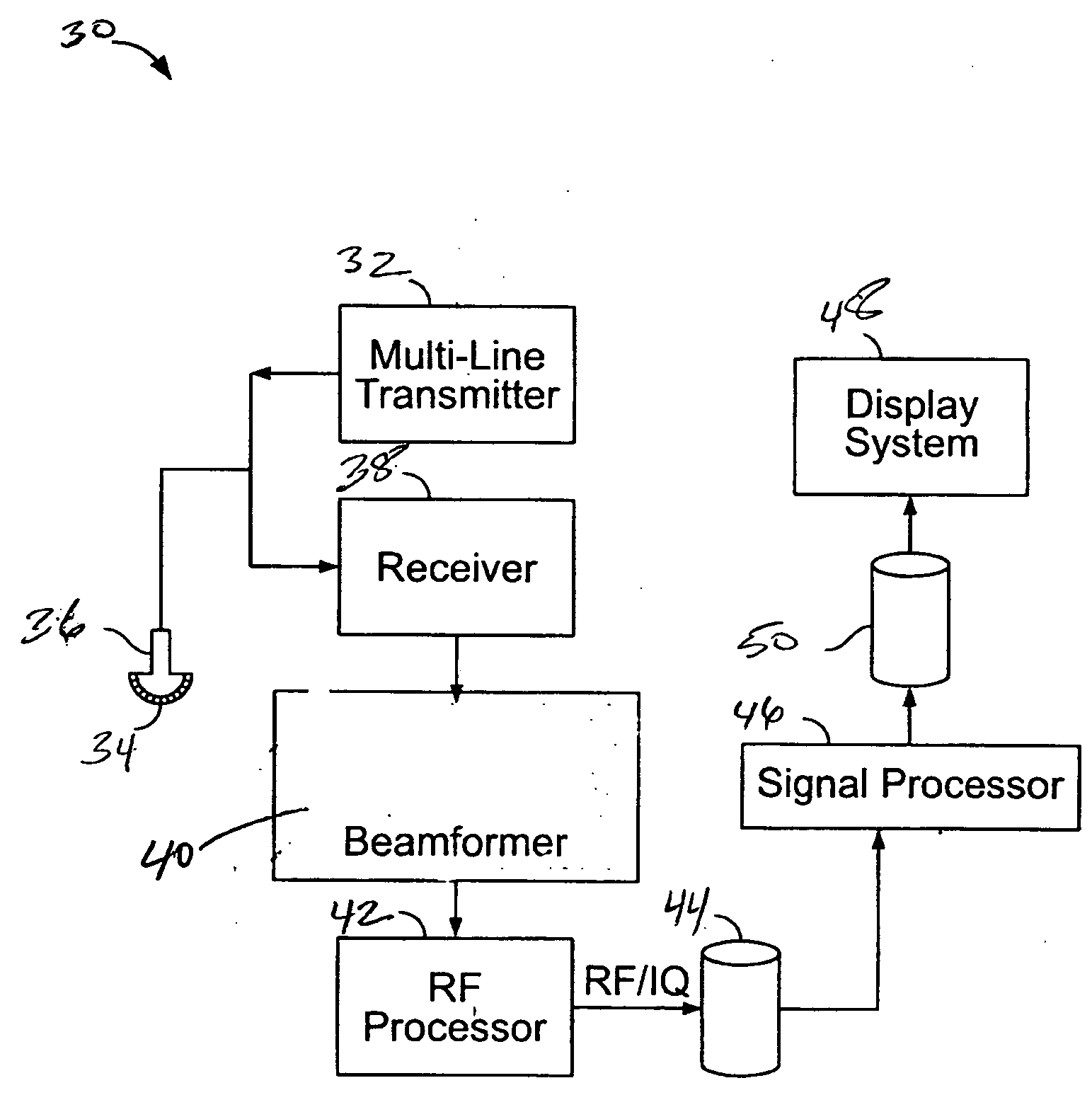
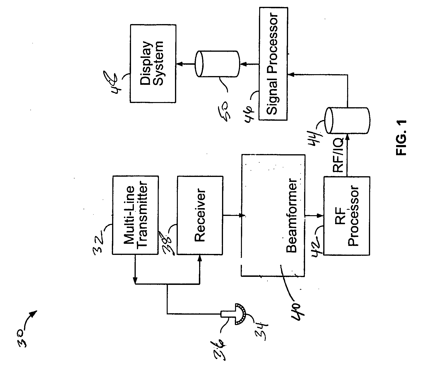
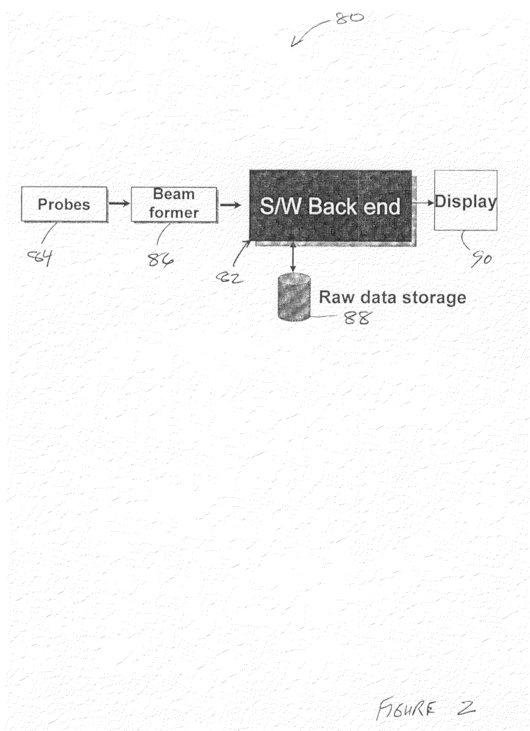
Method and systems for medical ultrasound imaging using a handheld ultrasound imaging system is provided. The ultrasound system includes a probe configured to acquire scan data from an object and a handheld device configured to process the received scan data and perform image compounding.

Description

BACKGROUND OF THE INVENTION[0001]This invention relates generally to ultrasound systems, and more particularly, handheld and hand-carried ultrasound (or other medical imaging) systems.[0002]Ultrasound imaging is a medical imaging technique for imaging organs and soft tissues in a human body. Ultrasound imaging uses real time, noninvasive high frequency sound waves to produce a two-dimensional (2D) image. Although, ultrasound imaging provides less anatomical information compared to CT or MRI, ultrasound imaging has several advantages in that patients are not exposed to radiation, studies of moving structures may be provided in real-time, the image scan is quickly performed and is inexpensive.[0003]In conventional ultrasound imaging, the image is acquired by a series of parallel scan lines. This results in an image in which some anatomical structures may be “shadowed” by objects closer to the transducer and diagonal structures may not be optimally imaged. Typically, when the boundarie...

Claims


Application Information

Patent Timeline
IPC IPC(8): A61B8/00
CPCA61B8/13G01S7/52047G01S15/8995G01S7/52085G01S7/52096G01S7/52066A61B8/4427A61B8/0841A61B8/5253
Inventor HALMANN, NAHI
Owner GENERAL ELECTRIC CO
Learn More