
Dilation introducer and methods for orthopedic surgery

a bone fixation device and introducer technology, applied in the direction of dilators, infusion syringes, applications, etc., can solve the problems of substantial trauma to the surrounding tissue, and achieve the effect of better observation of the procedur

Inactive Publication Date: 2008-11-20
INTERVENTIONAL SPINE
View PDF100 Cites 75 Cited by
  • Summary
  • Abstract
  • Description
  • Claims
  • Application Information

AI Technical Summary

Benefits of technology

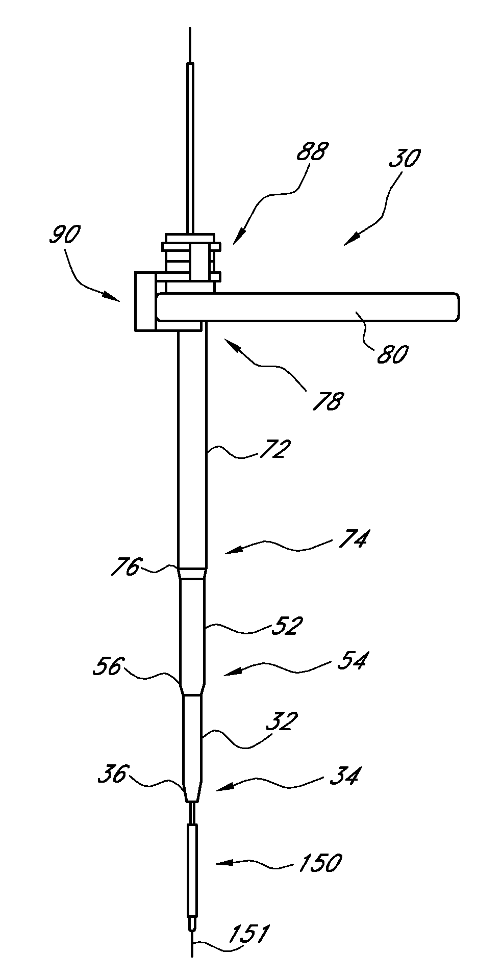
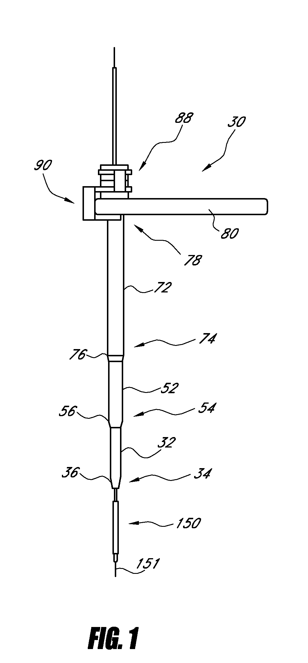
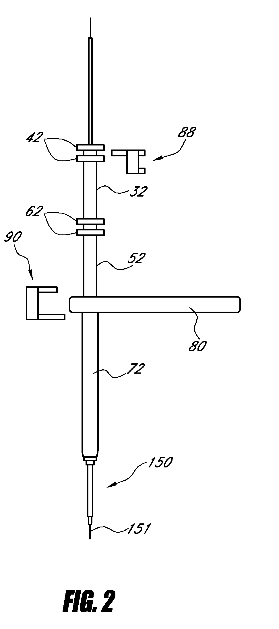
[0007]Briefly, and in general terms, the invention provides for a telescoping dilation introducer for orthopedic surgery, the dilation introducer having a locked assembled configuration for initial placement of the dilation introducer against a patient's tissue to be treated, and an unlocked, collapsed configuration for dilating the patient's soft surrounding tissue to a desired degree of dilation to permit minimally invasive surgical procedures on the patient's tissue. As the telescoping dilation introducer is inserted, each individual dilator tube is successively released and advanced to progressively expand the patient's soft tissue down to the tissue to be treated. In a particularly useful aspect of the invention, the tissue to be treated is bone tissue which must be prepared prior to attachment of adjacent bone section in a fusion process. While there are many applications of the dilation introducer of the invention, the invention is particularly applicable to fusion of bones in orthopedic surgery using minimally invasive technique, and will be described herein in particular applications of those procedures. The invention also concerns a minimally invasive procedure utilizing the telescoping dilation introducer to insert a bone fixation device into a patient's spine for posterior spine fusion. While posterior spine fusion currently takes up to two hours to complete, and requires a six inch incision, with the apparatus and method of the invention, comparable surgery can be completed in less than thirty minutes, with a dilation port 13 mm or less in diameter, thus lowering the chance of damage to the surrounding soft tissue.
[0008]The present invention accordingly provides for a dilation introducer for orthopedic surgery, the dilation introducer having a locked assembled configuration for initial placement of the dilation introducer against a patient's bone tissue to be treated, and an unlocked, collapsed configuration for dilating the patient's soft tissue to a desired degree of dilation to permit minimally invasive surgical procedures on the patient's bone tissue. The dilation introducer includes a first dilator tube having a distal end and a proximal end, a longitudinal lumen with a distal opening and a proximal opening, the distal end having a tapered tip, and a second dilator tube, the first dilator tube being removably received in the second dilator tube for slidable telescoping movement within the second dilator tube, the second dilator tube having a distal end and a proximal end, an inner lumen with a distal opening and a proximal opening, the distal end having a tapered tip. Alternatively, the first dilator tube may have a non-cannulated configuration, formed without a lumen. The dilation introducer also advantageously includes means for removably connecting the first and second dilator tubes together in a locked configuration, whereby in the locked configuration the distal end of the first dilator tube can be pressed against the patient's bone tissue to be treated, and whereby in the unlocked configuration the second dilator tube is permitted to slidably telescope over the first dilator tube to dilate the patient's soft tissue at the distal end of the dilation introducer.
[0021]In a further aspect of the invention, one or more of the dilation tubes may be fitted with light transmitting means, such as optional fibers and the like to illuminate the opening at the distal end of the dilation device to assist in visualizing the area where the procedure is being performed. Similarly, the dilation tubes can be fitted with imaging equipment to allow the surgeon to better observe the procedure being performed.

Problems solved by technology

In either type of procedure, there is substantial trauma to the surrounding tissue if a large incision is required.

Method used


Image

  • Dilation introducer and methods for orthopedic surgery
  • Dilation introducer and methods for orthopedic surgery
  • Dilation introducer and methods for orthopedic surgery

Examples


Embodiment Construction

[0059]Referring to the drawings, which are provided for purposes of illustration and by way of example, the present invention provides for a telescoping dilation introducer for orthopedic surgery, the dilation introducer having a locked assembled configuration for initial placement of the dilation introducer against a patient's bone tissue to be treated, and an unlocked, collapsed configuration dilating the patient's soft tissue down to the bone tissue to be treated to a desired degree of dilation to permit minimally invasive surgical procedures on the patient's bone tissue to be treated.

[0060]While the invention will be described with specificity to a spinal fusion procedure, those skilled in the art will recognize that the apparatus and method of the art will recognize that the apparatus and method of the invention can also be advantageously used for procedures in which the dilation introducer can be brought up against other firm or solid structures in the body or introduced into ...


PUM


Abstract

A method is provided for a minimally invasive procedure utilizing the telescoping dilation introducer to insert a bone fixation device into a patient's spine for posterior spine fusion. The dilation introducer has a locked assembled configuration for placement of the dilation introducer against a patient's bone tissue to be treated, and an unlocked, collapsed configuration for dilating the patient's soft tissue down to the bone tissue to be treated to a desired degree of dilation to permit minimally invasive surgical procedures on the patient's bone tissue to be treated. Dilator tubes are successively released and advanced to progressively expand the patient's soft tissue down to the bone tissue to be treated.

Description

CROSS-REFERENCE TO RELATED APPLICATIONS[0001]This application is a divisional of U.S. application Ser. No. 10 / 911,215, filed on Aug. 3, 2004, the entire contents of which is hereby incorporated herein by reference.BACKGROUND OF THE INVENTION[0002]1. Field of the Invention[0003]This invention relates to bone fixation devices, and more particularly relates to a dilation introducer for introducing a bone fixation device for orthopedic surgery, such as for vertebral fusion.[0004]2. Description of the Related Art[0005]Fusion of two adjacent vertebrae is a common surgical treatment for back injuries due to damage or defects in a spinal disc between two adjacent vertebrae, such as conditions due to a herniated disc or disc degeneration. The entire disc may be removed by a discectomy procedure, and may be replaced with bone or a bone substitute and / or cage in order to prevent collapse of the disc space between the adjacent vertebrae. Early techniques for stabilizing the adjacent vertebrae i...

Claims


Application Information

Patent Timeline
Patent Type & Authority Applications(United States)
IPC IPC(8): A61B19/00A61M19/00A61M5/178
CPCA61B17/02A61B17/3417A61B17/3421A61B2017/00477A61B2017/320044A61M29/00
Inventor CULBERT, BRAD S.BOOMER, MARK C.WARREN, CHRISTOPHER
Owner INTERVENTIONAL SPINE
Learn More