Image acquisition, archiving and rendering system and method for reproducing imaging modality examination parameters used in an initial examination for use in subsequent radiological imaging

a technology of image acquisition and image archiving, applied in tomography, applications, instruments, etc., can solve the problems of inability to precisely repeat initial examinations, occupied by patients, ct or mrt apparatuses,

Inactive Publication Date: 2008-12-18
SIEMENS HEALTHCARE GMBH
View PDF7 Cites 41 Cited by
  • Summary
  • Abstract
  • Description
  • Claims
  • Application Information

AI Technical Summary

Benefits of technology

[0013]An object of the present invention is to allow image data of slice exposures acquired in the framework of an initial examination by means of CT-assisted or MRT-assisted imaging and / or 2D projections or, respectively, 3D views of specific tissue regions, internal organs, anatomical subjects or, respectively, pathological structures inside the body of a patient to be examined (which 2D projections or 3D views are reconstructed in the framework of a subsequent image post-processing process) to be reproduced in following monitoring examinations more exactly than has previously been possible.

Problems solved by technology

A further problem of present CT-assisted or MRT-assisted imaging, image archiving and image processing systems is that the position occupied by a patient on the examination table of a CT or MRT apparatus (i.e. the patient position) in an initial examination cannot be exactly repeated upon implementation of a follow-up examination, i.e. with the same position and angle coordinates relative to a three-dimensional Cartesian coordinate system I (patient coordinate system) which is spanned by the longitudinal axis z of the body of the patient and two direction vectors proceeding orthogonal to one another in the x-direction (transverse-horizontal) or in the y-direction (transverse-vertical).
This is a relatively time-consuming process that must be manually executed by a radiologist implementing both examinations.
Moreover, the spatial orientation of the slices shown in an initial examination in the framework of an MRT-assisted imaging process, nor the spatial orientation of 2D or 3D reconstructions that were generated in the framework of a post-processing process implemented after the initial examination, are not reproducible in commercially available CT and MRT systems.
Rather, such information must be estimated on the basis of the respective prior examination and be manually input by the radiologist conducting the examinations, which (apart from the not inconsiderable time expenditure that accumulates over multiple follow-up examinations) is prone to a relatively large imprecision of the image reproduction.

Method used


Image

  • Image acquisition, archiving and rendering system and method for reproducing imaging modality examination parameters used in an initial examination for use in subsequent radiological imaging
  • Image acquisition, archiving and rendering system and method for reproducing imaging modality examination parameters used in an initial examination for use in subsequent radiological imaging
  • Image acquisition, archiving and rendering system and method for reproducing imaging modality examination parameters used in an initial examination for use in subsequent radiological imaging

Examples


Embodiment Construction

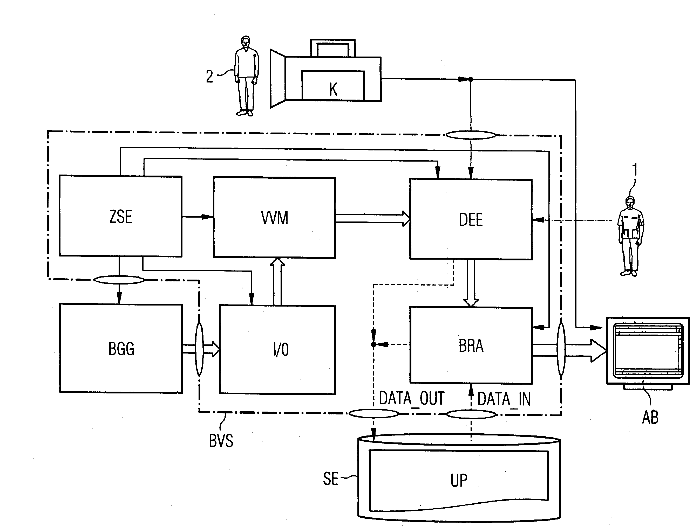
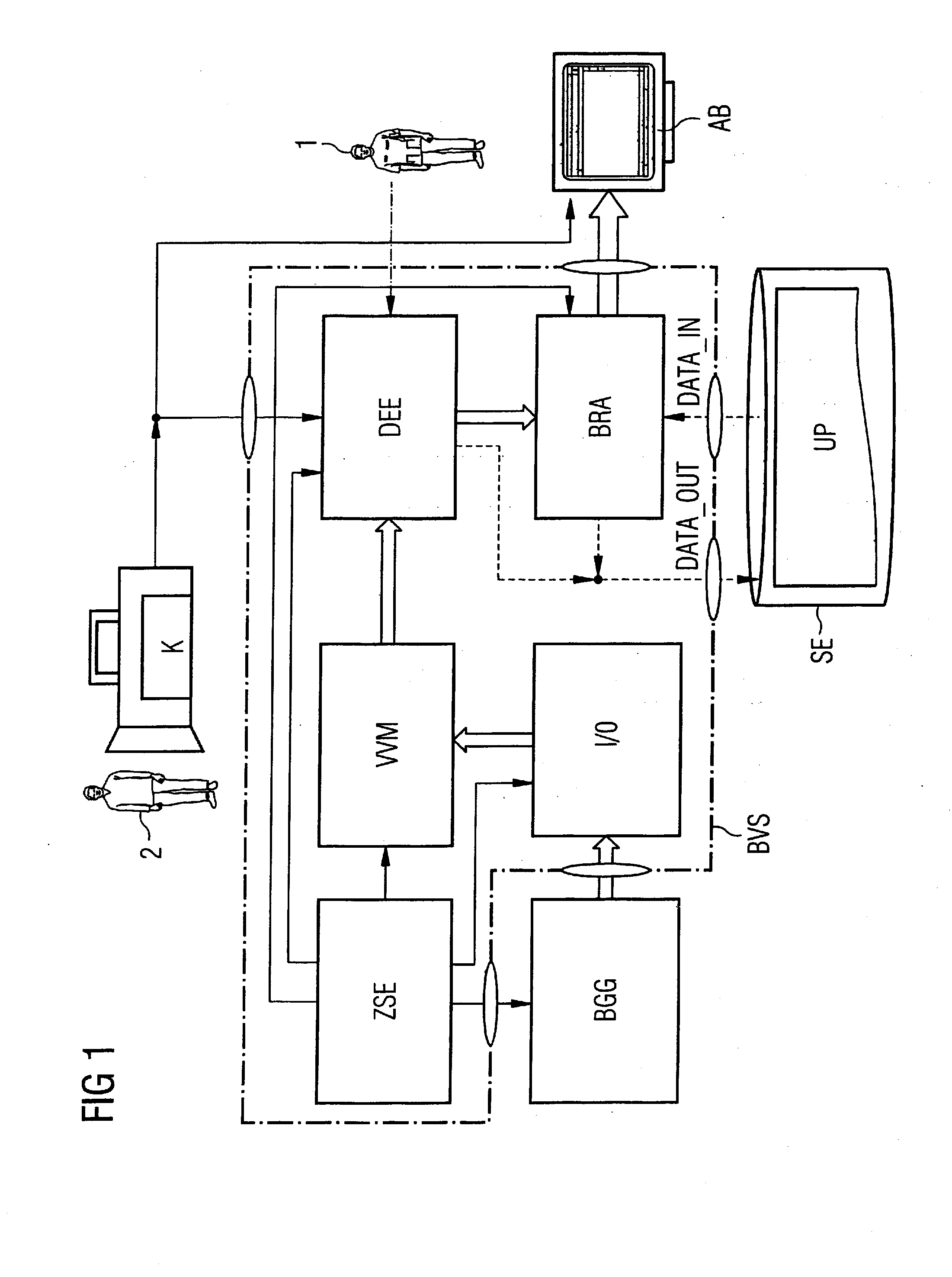
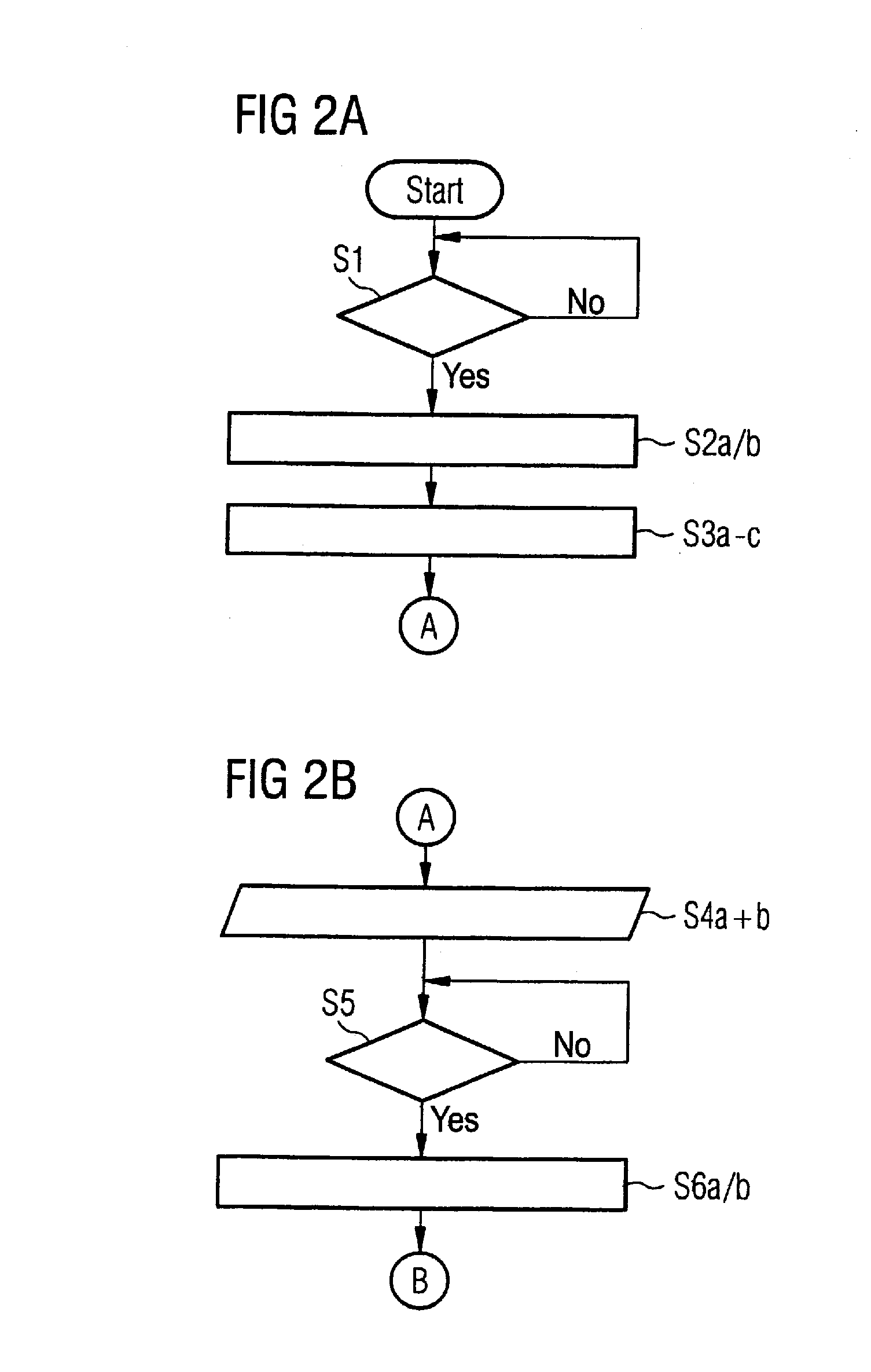
[0041]The system components of the image acquisition, image archiving and image rendering system according to the invention and the steps of the associated method according to the invention are described in detail in the following.

[0042]A schematic block diagram of an embodiment of an image acquisition, image archiving and image rendering system according to the present invention is shown in FIG. 1. This system enables image data (generated by a medical technology imaging apparatus BGG (for example by a CT or MRT apparatus)) of tissue regions inside the body of a patient 2 to be examined to be displayed in the form of slice exposures or in the form of reconstructed 2D projections or reconstructed 3D presented from arbitrary projection angles. The image data provided by a computed or magnetic resonance tomography imaging modality are supplied to an image processing system BVS via an input / output interface I / O. In addition to a central processing device ZSE which controls the data exc...


PUM


Abstract

A CT- or MRT-assisted image acquisition, image archiving and image rendering system allows generation, storage, post-processing, retrieval and graphical visualization of computed or magnetic resonance tomography image data that, for example, can be used in the clinical field in the framework of radiological slice image diagnostics as well as in the framework of interventional radiology. Moreover, a method implemented by this system allows reproduction of patient-specific examination parameters of an initial examination implemented by means of computed or magnetic resonance tomography imaging in the framework of CT or MRT follow-up examinations (“follow-ups”), for example in a post-operative tumor examination implemented under slice image monitoring in connection with a histological tissue sample extraction (biopsy) implemented under local anesthesia or a minimally-invasive intervention implemented for tumor treatment. Acquisition, measurement, 2D and / or 3D reconstruction parameters from a radiological initial examination of the patient conducted by means of CT, PET-CT or MRT as well as position data to establish the position adopted by this patient on the examination table of a computed tomography or magnetic resonance tomography apparatus are electronically documented, retrievably and persistently stored, and are automatically reused given subsequent CT-, PET-CT- or MRT-based monitoring examinations, or CT-controlled or MRT-controlled interventional or operative procedures.

Description

BACKGROUND OF THE INVENTION[0001]1. Field of the Invention[0002]The present invention concerns a CT-assisted or MRT-assisted image acquisition, image archiving and image rendering system for generation, storage, post-processing, retrieval and graphical visualization of computed or magnetic resonance tomography image data that, for example, can be used in the clinical field in the framework of radiological slice image diagnostics as well as in the framework of interventional radiology. The present invention also concerns a method implemented by such a system for reproduction of patient-specific examination parameters of an initial examination implemented by means of computer or magnetic resonance tomography imaging in the framework of CT or MRT follow-up examinations (“follow-ups”), for example in a post-operative tumor examination implemented under slice image monitoring in connection with a histological tissue sample extraction (biopsy) implemented under local anesthesia or a minim...

Claims


Application Information

Patent Timeline
Patent Type & Authority Applications(United States)
IPC IPC(8): G06K9/00G16H30/20
CPCA61B6/032A61B6/545G06F19/321G16H30/20G16H40/63
Inventor BOEING, DIETERENGELS, HEINZ-PETER
Owner SIEMENS HEALTHCARE GMBH
Why Eureka
Try Eureka