Method and system for detection of obstructions in vasculature

a vasculature and obstruction technology, applied in the field of medical imaging examination methods and apparatuses, can solve the problems of reducing the detection accuracy of vasculature,

Inactive Publication Date: 2009-01-08
GENERAL ELECTRIC CO
View PDF20 Cites 140 Cited by
  • Summary
  • Abstract
  • Description
  • Claims
  • Application Information

AI Technical Summary

Problems solved by technology

However, when a large artery in the vasculature is blocked by blood clots or other particles, the amount of blood supplied to the tissue may be insufficient, eventually causing tissue to die.
For example, the presence of plaque restricts blood flow, damages the blood vessel wall and and / or promotes blood clot formation.
In addition, calcification of blood vessels occurs where lipids accumulate in the blood vessel wall leading to hardening of the arteries.
Also, an embolus in a blood vessel may cause severe dysfunction of an organ, which may be fatal if undiagnosed and / or untreated.
For instance, blockage of the internal carotid artery may reduce blood supply to the brain, causing a stroke.
However, when a pulmonary artery is blocked by an embolus, plaque or calcification, the amount of blood supplied to the lung tissue may be insufficient, eventually causing tissue to die.
Pulmonary embolism is one of the leading causes of sudden deaths in hospitalized patients.
Also, symptoms associated with pulmonary emboli may include difficulty breathing, pain during breathing, and more rarely circulatory instability and death.
Unfortunately, the diagnosis is often missed because patients with pulmonary emboli present nonspecific signs and symptoms.
The considerable amount of data associated with a single CT scan presents a time-consuming process to the radiologist.
Furthermore, this substantial amount of data may disadvantageously lead to missed detection of pulmonary emboli, as it is difficult to identify a suspicious area in an extensive amount of data.
However, detection of pulmonary emboli in these relatively small vessels is a challenging task.
In addition, confusion caused by branching points in the vasculature, veins, motion artifacts, partial volume and other pathologies may lead to misdiagnosis.
However, one of the vital but complex problems encountered by CAD schemes is the initial selection of potential thresholds.
However, traditional methods of thresholding the CT images encounter problems associated with the varying contrast levels that are dependent on the branching level that make the proper selection of a threshold very difficult, thereby resulting in an elevated number of misdiagnosis, and consequently requiring additional radiologist time.
More particularly, a single global intensity threshold may not be able to separate the entire vessel tree.
Selecting a tight threshold bandwidth may result in under-segmentation of smaller branches, while selecting a loose threshold bandwidth may result in inclusion of background structures such as airway walls and interstitial lung disease, if present.

Method used


Image

  • Method and system for detection of obstructions in vasculature
  • Method and system for detection of obstructions in vasculature
  • Method and system for detection of obstructions in vasculature

Examples


Embodiment Construction

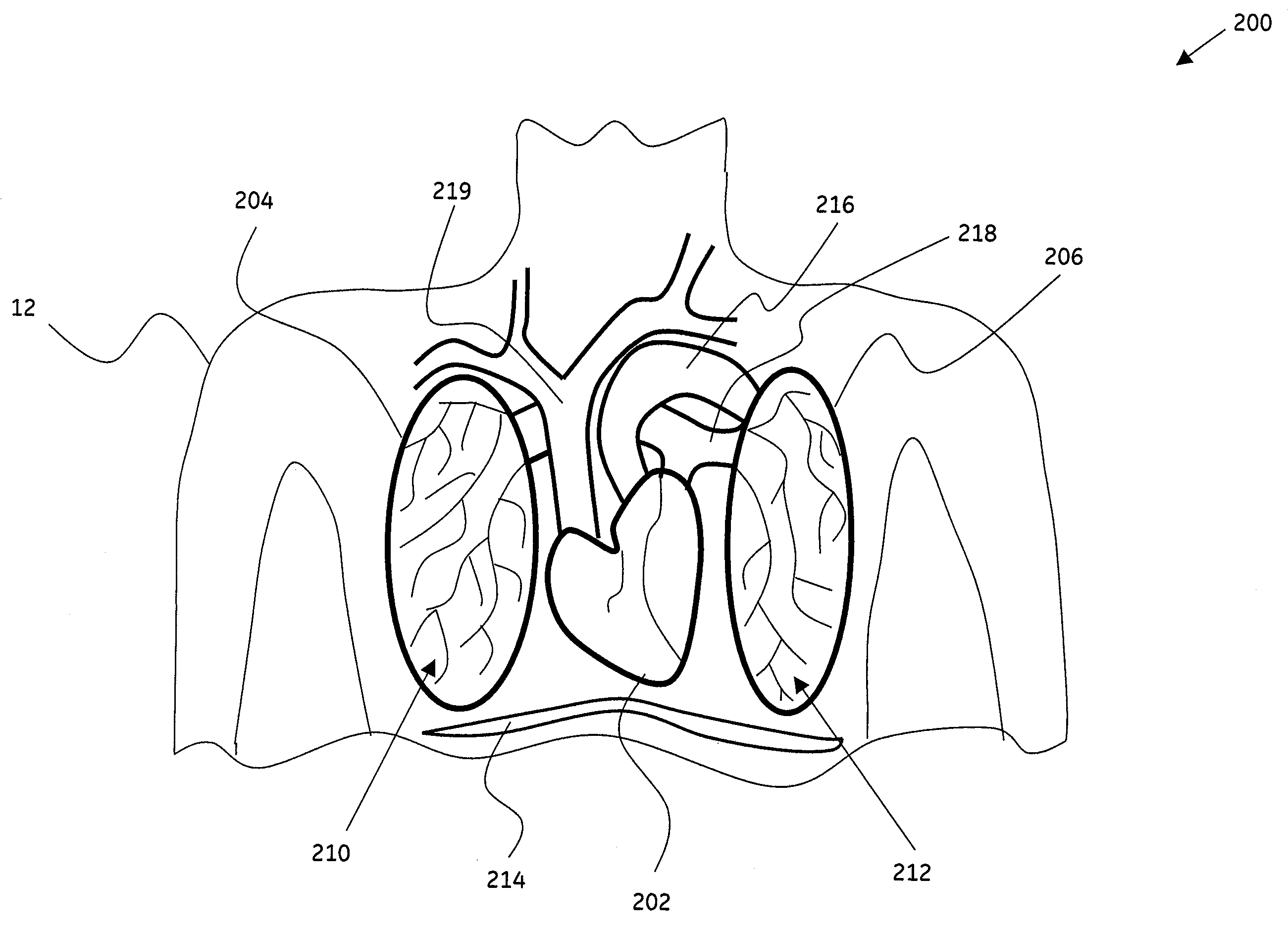
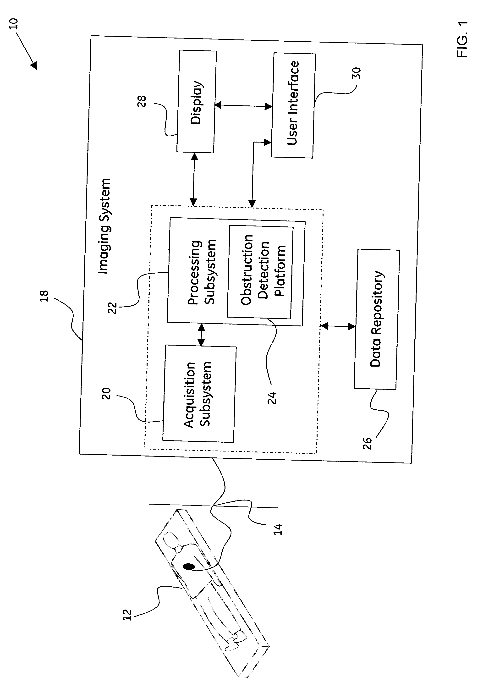
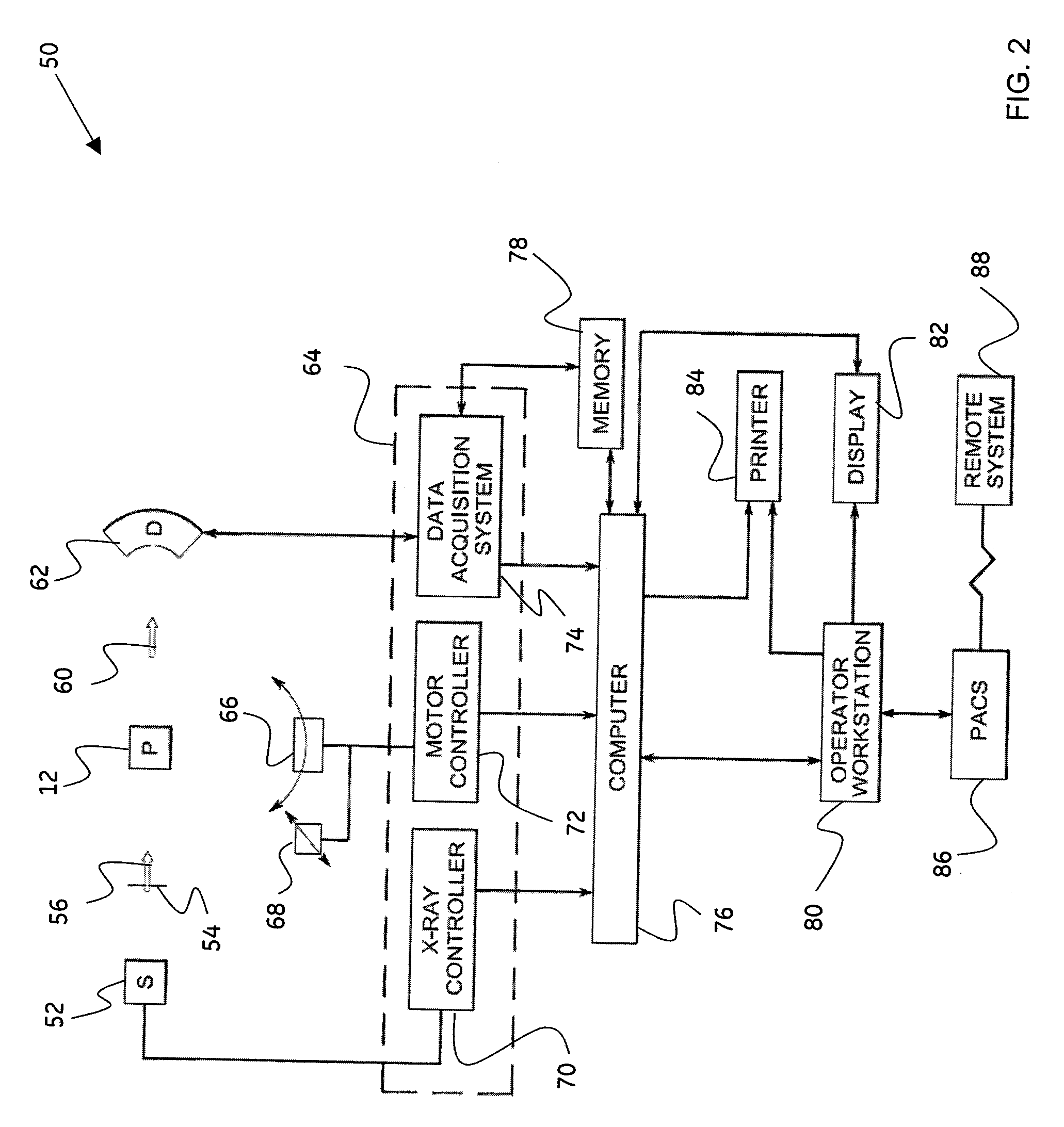
[0023]As will be described in detail hereinafter, a method for automatic detection of obstructions in vasculature and a system for automatic detection of obstructions in vasculature configured to optimize detection of obstructions in vasculature and simplify clinical workflow in a diagnostic imaging system, are presented. Employing the method and system described hereinafter, the system for the automatic detection of obstructions may be configured to facilitate substantially superior detection of obstructions in vasculature, thereby simplifying the clinical workflow of the detection of obstructions.

[0024]Although, the exemplary embodiments illustrated hereinafter are described in the context of a medical imaging system, it will be appreciated that use of the diagnostic system in industrial applications are also contemplated in conjunction with the present technique.

[0025]FIG. 1 is a block diagram of an exemplary system 10 for use in diagnostic imaging in accordance with aspects of t...


PUM


Abstract

A method for automatic detection of obstructions in vasculature in an anatomical region is presented. The method includes partitioning the anatomical region into a plurality of sub-regions based at least in part on anatomical knowledge. Further, the method includes adaptively computing a threshold intensity value corresponding to each of the plurality of sub-regions. Additionally, the method includes extracting the vasculature in each of the plurality of sub-regions based on the corresponding computed threshold intensity value, where the extracted vasculature comprises a plurality of vessel segments. The method also includes detecting an obstruction in the extracted vasculature. Systems and computer-readable medium that afford functionality of the type defined by this method is also contemplated in conjunction with the present technique.

Description

BACKGROUND[0001]The invention relates generally to methods and apparatus for review of medical imaging exams, and more particularly to methods and apparatus for review of image data, such as that resulting from lung exams.[0002]Blood vessels in the vasculature typically aid in the transportation of blood to various parts of the body. However, when a large artery in the vasculature is blocked by blood clots or other particles, the amount of blood supplied to the tissue may be insufficient, eventually causing tissue to die. For example, the presence of plaque restricts blood flow, damages the blood vessel wall and and / or promotes blood clot formation. In addition, calcification of blood vessels occurs where lipids accumulate in the blood vessel wall leading to hardening of the arteries. Also, an embolus in a blood vessel may cause severe dysfunction of an organ, which may be fatal if undiagnosed and / or untreated. For instance, blockage of the internal carotid artery may reduce blood s...

Claims


Application Information

Patent Timeline
Patent Type & Authority Applications(United States)
IPC IPC(8): A61B6/00
CPCA61B5/02007A61B5/055A61B5/489G06T2207/30101A61B6/481A61B6/504G06T7/0012A61B6/032
Inventor DUTTA, SANDEEPGOPINATH, AJAYSURYANARAYANAN, SRIKANTHROY, ARUNABHA SHASANKAMENDONCA, PAULO RICARDO DOS SANTOS
Owner GENERAL ELECTRIC CO
Why Eureka
Try Eureka