Ultrasonic image processing apparatus and a method for processing an ultrasonic image

a processing apparatus and ultrasonic image technology, applied in the field of ultrasonic image, can solve the problems of difficult to properly evaluate the wall motion at each part of the myocardium, the direction of the myocardium represented, and the shape of the cardiac cavity represented in an image on the plane significantly different from the actual shap

Active Publication Date: 2009-03-05
TOSHIBA MEDICAL SYST CORP
View PDF3 Cites 30 Cited by
  • Summary
  • Abstract
  • Description
  • Claims
  • Application Information

AI Technical Summary

Benefits of technology

[0013]In a first aspect of the present invention, an ultrasonic image processing apparatus comprises: a contour specifying part configured to receive volume data representing a subject acquired by transmission of ultrasonic waves to the subject and specify a 3-dimensional contour of a myocardium based on the volume data; a forming part configured to set a reference point...

Problems solved by technology

Therefore, when the wall motion information is superimposed and displayed on the myocardium represented in the short-axis image according to the conventional technique, a problem arises in which the thickness direction of the myocardium represented in the short-axis image does not coincide with the direction of the vector in the actual wall-thickness direction of the myocardium.
Therefore, in the conventional technique, it is difficult to properly evaluate a wall motion at each part of the myocardium, even if observing the morphology and wall motion information of the myocardium represented in the short-axis image.
Further, according to the method of setting a plane from the central axis toward the myocardium, there is a problem that, depending on the angle of the plane set from the central axis toward the myocardium...

Method used


Image

  • Ultrasonic image processing apparatus and a method for processing an ultrasonic image
  • Ultrasonic image processing apparatus and a method for processing an ultrasonic image
  • Ultrasonic image processing apparatus and a method for processing an ultrasonic image

Examples


first modification

(First Modification)

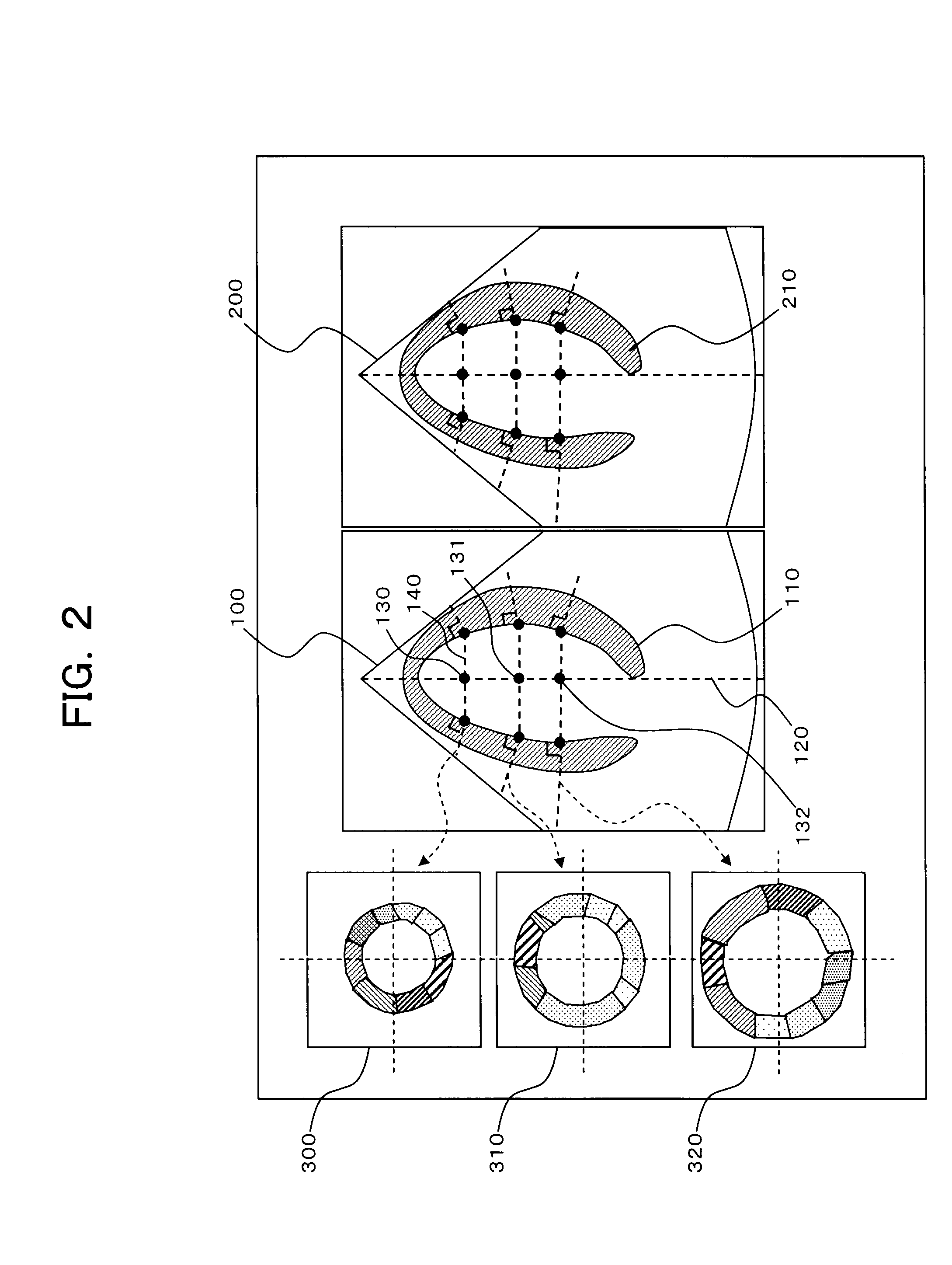
[0143]Next, a first modification of the ultrasonic imaging apparatus according to the abovementioned embodiment will be described with reference to FIGS. 7, 8A and 8B. FIG. 7 is a view of a screen illustrating an example of an image displayed on the display in the first modification. FIGS. 8A and 8B are schematic views illustrating the contour of the myocardium on a cross-section along the long-axis direction of the myocardium.

[0144]In the first modification, the image-generation-plane forming part 77 forms an image generation plane by executing a different process from that in the abovementioned embodiment. Each part other than the image-generation-plane forming part 77 executes the same process as in the abovementioned embodiment.

[0145]Similarly in the abovementioned embodiment, the image generator 6 reads out volume data correlated with the cardiac phase designated by the operator from the storage 5, and generates long-axis image data of a long-axis cross-sect...

second modification

(Second Modification)

[0178]Next, a second modification of the ultrasonic imaging apparatus 1 according to the abovementioned embodiment will be described with reference to FIG. 10A and FIG. 10B. FIG. 10A and FIG. 10B are schematic views illustrating the contour of the myocardium in a cross-section along the short-axis direction of the myocardium.

[0179]In this modification 2, the second-plane forming part 76 executes a process different from that in the above-mentioned embodiment, thereby forming second planes orthogonal to the myocardium.

[0180]First, the operator designates an arbitrary cardiac phase by using the operation part 12. The cardiac phase designated by the operator is set as the initial time phase. Further, the operator designates an arbitrary cross-section for the volume data by using the operation part 12. In this second modification, the operator designates a short-axis cross-section of the heart. Information showing the initial time phase designated by the operator an...


PUM


Abstract

A contour specifying part receives volume data representing a subject acquired by transmission of ultrasonic waves to the subject, and specifies a 3-dimensional contour of a myocardium based on the volume data. A forming part sets a reference point on the contour of the myocardium, and forms an image generation plane including a plane substantially orthogonal to the contour of the myocardium at the reference point. An image generator generates image data on the image generation plane based on the volume data. A display controller controls a display to display an image based on the image data.

Description

BACKGROUND OF THE INVENTION[0001]1. Field of the Invention[0002]The present invention relates to an ultrasonic image processing apparatus that obtains the motion state of a subject by using an ultrasonic image of the subject acquired with ultrasonic waves, and also relates to a method for processing an ultrasonic image.[0003]2. Description of the Related Art[0004]Objective and quantitative evaluation of functions of body tissues such as the myocardium of a heart is very important for diagnosis of the body tissues. For example, a method of acquiring image data of a heart with an ultrasonic imaging apparatus and quantitatively evaluating based on the image data has been proposed.[0005]For example, a method of executing a pattern matching process on images and obtaining motion information such as displacement and strain of tissues has been proposed. As a specific example, for evaluation of the myocardium of a heart, ultrasonic waves are transmitted to the heart, and volume data that re...

Claims


Application Information

Patent Timeline
IPC IPC(8): A61B8/00
CPCA61B8/0858A61B8/0883A61B8/14A61B8/463A61B8/483A61B8/523G01S15/8993G01S7/52063G01S7/52071G01S7/52073G01S7/52074G01S7/52088G01S15/8979G01S7/52042
Inventor OHUCHI, HIROYUKIKAWAGISHI, TETSUYAABE, YASUHIKONISHIURA, MASAHIDETAKEGUCHI, TOMOYUKI
Owner TOSHIBA MEDICAL SYST CORP
Why Eureka
Try Eureka