Method and system for polyp segmentation for 3D computed tomography colonography

a computed tomography and colonography technology, applied in image enhancement, image analysis, instruments, etc., can solve the problems of large variability in physician's polyp measurement, difficult polyp segmentation, and unsupervised approaches that work well

Inactive Publication Date: 2009-03-19
SIEMENS MEDICAL SOLUTIONS USA INC
View PDF13 Cites 33 Cited by
  • Summary
  • Abstract
  • Description
  • Claims
  • Application Information

AI Technical Summary

Problems solved by technology

For example, a polyp may be hidden in a colonic fold and thus could be missed during the physician's visual inspection.
However, there is a large variability in physician's measurements of polyps.
Polyp segmentation is a challenging task because polyps are abnormal growths from the colon wall and the “expected” segmentations are often a semantic, perceptual boundary with low imaging contrast support.
These unsupervised approaches work well for up to 70% of polyps due to unclear polyp / nonpolyp boundaries, large within-class polyp appearance / shape variations, or limited heuristic shape assumptions.

Method used


Image

  • Method and system for polyp segmentation for 3D computed tomography colonography
  • Method and system for polyp segmentation for 3D computed tomography colonography
  • Method and system for polyp segmentation for 3D computed tomography colonography

Examples


Embodiment Construction

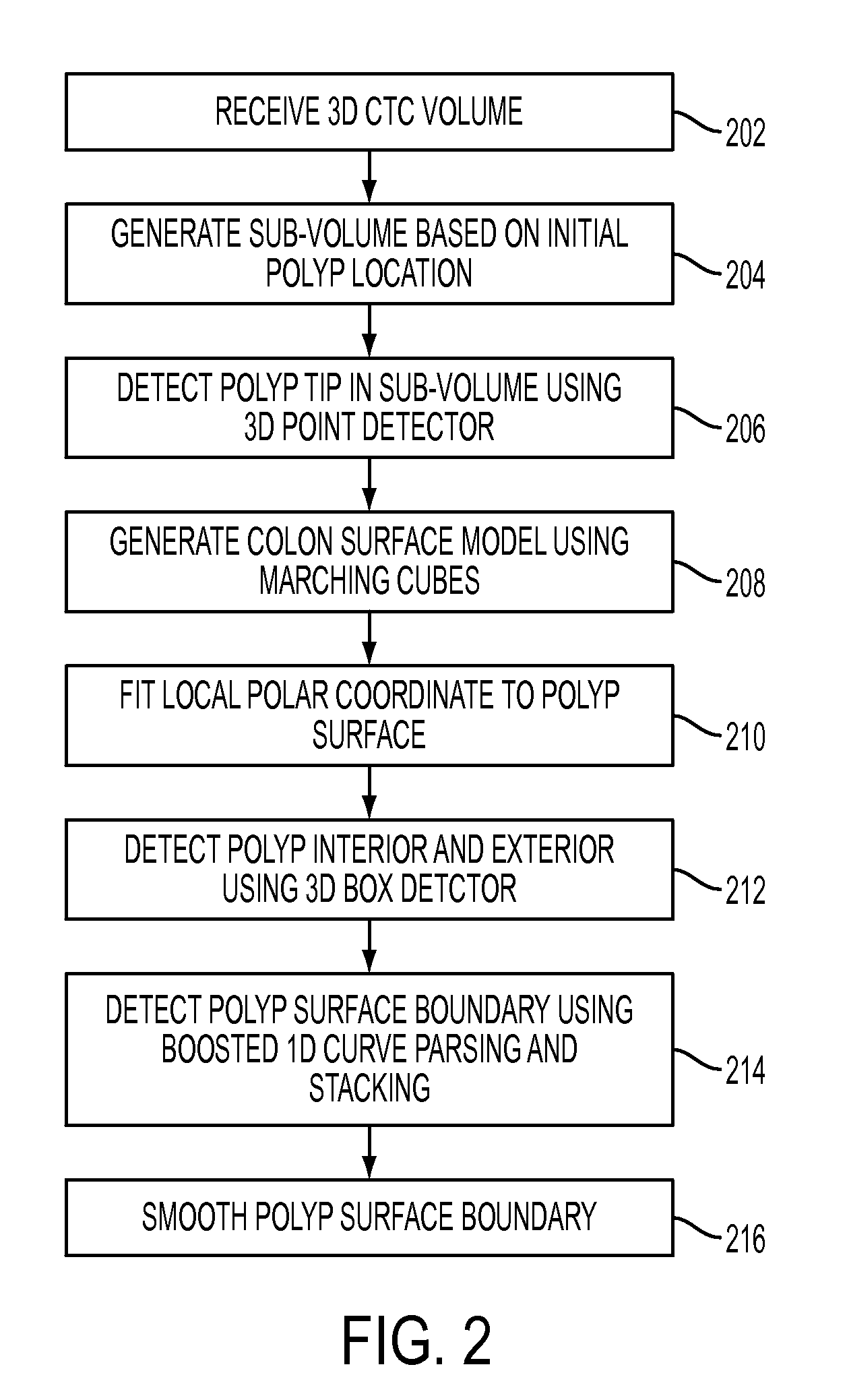
[0017]The present invention is directed to a method for polyp segmentation in 3D computed tomography colonography (CTC) volumes. Embodiments of the present invention are described herein to give a visual understanding of the polyp segmentation method. A digital image is often composed of digital representations of one or more objects (or shapes). The digital representation of an object is often described herein in terms of identifying and manipulating the objects. Such manipulations are virtual manipulations accomplished in the memory or other circuitry / hardware of a computer system. Accordingly, is to be understood that embodiments of the present invention may be performed within a computer system using data stored within the computer system.

[0018]Polyp segmentation, as performed in embodiments of the present invention, is the extraction of a polyp from a CTC volume given an initial position. The initial position can be manually selected by a physician or output by computer aided d...


PUM


Abstract

A method and system for polyp segmentation in computed tomography colonogrphy (CTC) volumes is disclosed. The polyp segmentation method utilizes a three-staged probabilistic binary classification approach for automatically segmenting polyp voxels from surrounding tissue in CTC volumes. Based on an input initial polyp position, a polyp tip is detected in a CTC volume using a trained 3D point detector. A local polar coordinate system is then fit to the colon surface in the CTC volume with the origin at the detected polyp tip. Polyp interior voxels and polyp exterior voxels are detected along each axis of the local polar coordinate system using a trained 3D box. A boundary voxel is detected on each axis of the local polar coordinate system based on the detected polyp interior voxels and polyp exterior voxels by boosted 1D curve parsing using a trained classifier. This results in a segmented polyp boundary.

Description

[0001]This application claims the benefit of U.S. Provisional Application Ser. No. 60 / 973,497, filed Sep. 19, 2007, the disclosure of which is herein incorporated by reference.BACKGROUND OF THE INVENTION[0002]The present invention relates to 3D computed tomography (CT) colonography, and more particularly, to automated polyp segmentation for 3D CT colonography.[0003]Colon cancer is the number two cause of cancer death in men and women combined, but it is one of the most preventable cancers because doctors can identity and remove pre-cancerous growths known as polyps. 3D CT colonography (CTC), or virtual colonoscopy, is emerging as a powerful polyp screening tool because of its non-invasiveness, low cost, and high sensitivity. 3D CTC visualizes the colon, and allows a physician to navigate the colon to search for polyps. However, the physician needs to manually adjust the navigation speed and change the angle in order to see a polyp clearly. For example, a polyp may be hidden in a col...

Claims


Application Information

Patent Timeline
Patent Type & Authority Applications(United States)
IPC IPC(8): G06K9/00
CPCG06T2207/20076G06T2207/30032G06T2207/10081G06T7/0081G06T7/0087G06T7/0012G06T7/11G06T7/143
Inventor LU, LEBARBU, ADRIANWOLF, MATTHIASLAKARE, SARANGBOGONI, LUCASALGANICOFF, MARCOSCOMANICIU, DORIN
Owner SIEMENS MEDICAL SOLUTIONS USA INC
Why Eureka
Try Eureka