

Composition and method of treating temporary and permanent hearing loss
- Summary
- Abstract
- Description
- Claims
- Application Information
AI Technical Summary
Benefits of technology
Problems solved by technology
Method used
Image

Examples
Example
Comparative Example 1
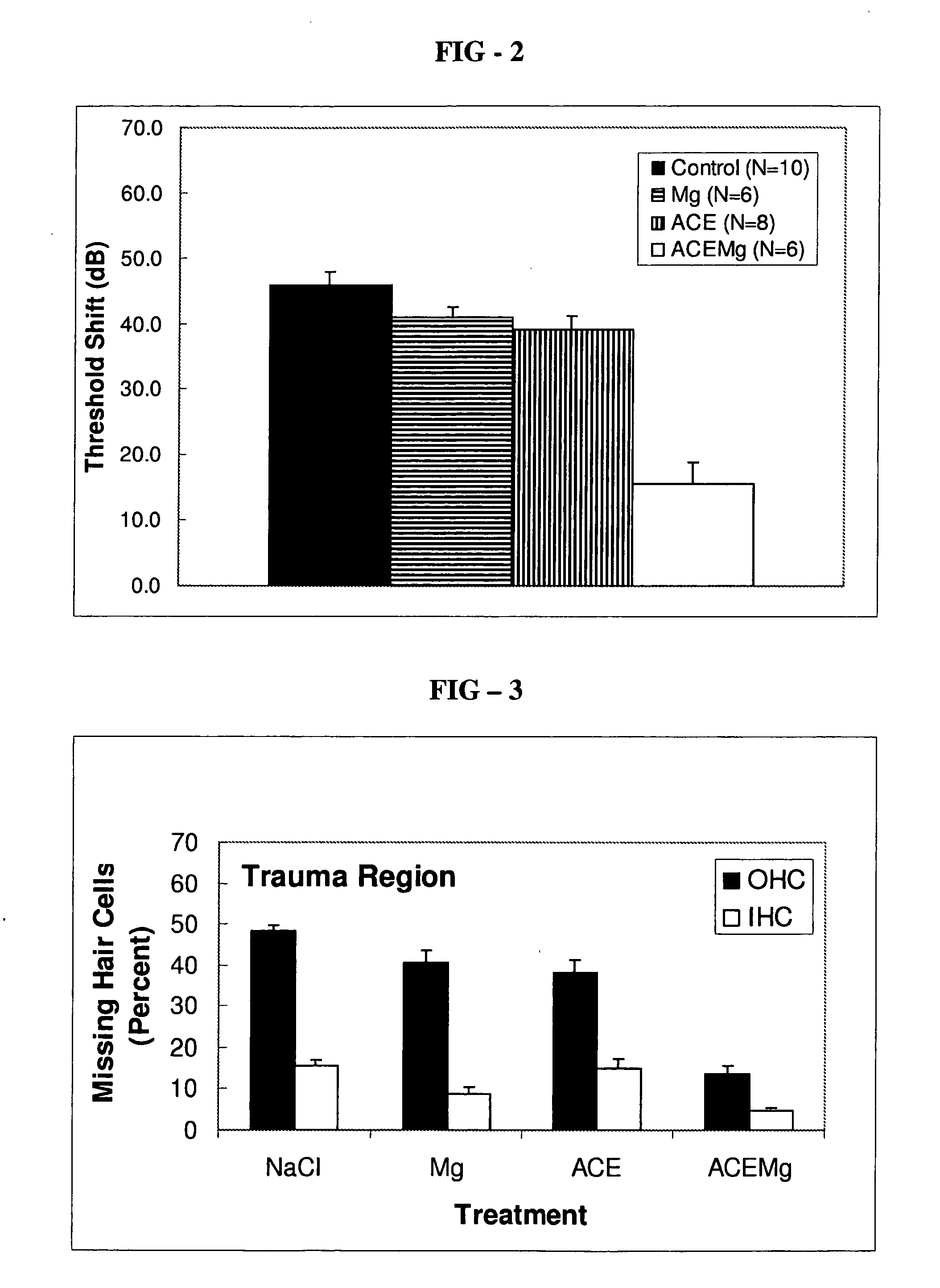
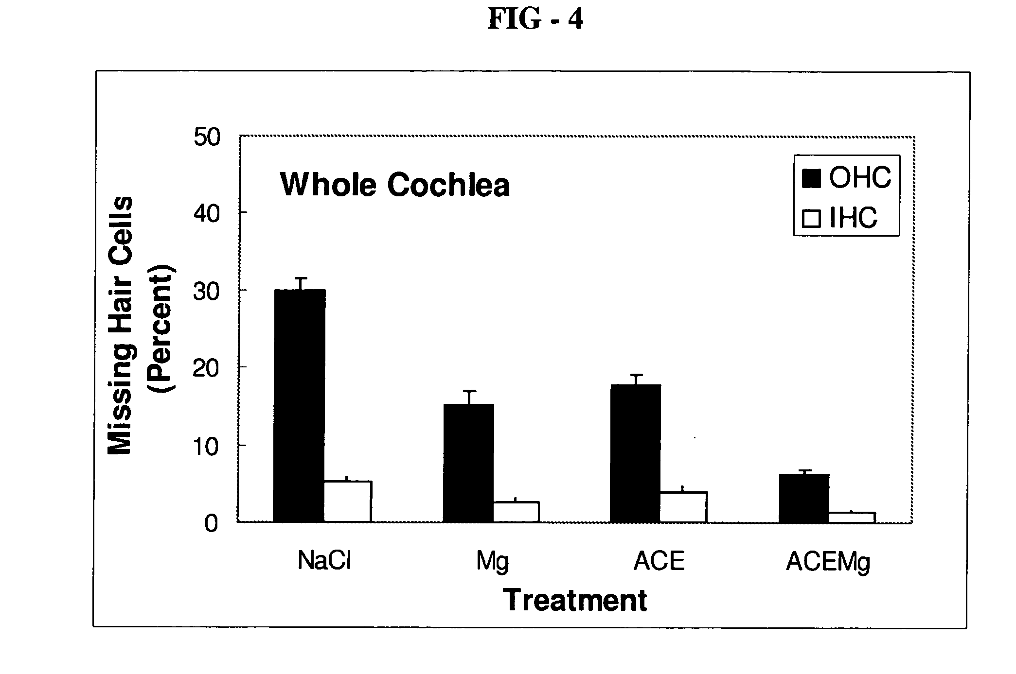
[0086]Guinea pigs are treated with other compositions in order to compare the efficacy of the composition of the present invention with the other compositions. For example, guinea pigs are separately treated in the same way as specified above in the Example with the following compositions: a saline (NaCl) composition as a control, a composition including only magnesium sulfate (2.85 mmol / kg) as the active ingredient, or a composition including only vitamins A (2.1 mg / kg beta-carotene), C (71.4 mg / kg ascorbic acid), and E (26 mg / kg Trolox®) (“ACE”) as the active ingredients. The guinea pigs are subjected to the same ABR testing, and the components of the ear are dissected, as described above in the Example to provide information on threshold shift and hair cell loss. Threshold shift and hair cell loss resulting from treatment with the other compositions are shown in FIGS. 2-4. After treatment according to the method of the present invention, outer hair cell loss ...
Example
Comparative Example 2
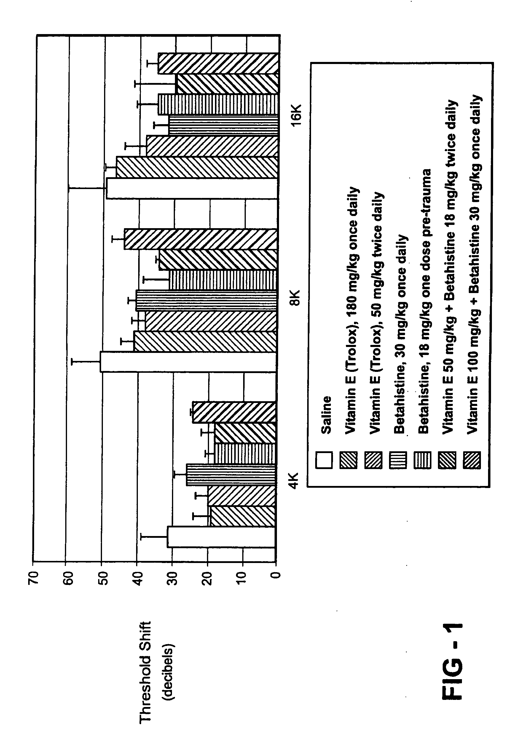
[0087]Guinea pigs are treated with a composition of vitamin E alone, betahistine alone, or a combination of betahistine and vitamin E to determine if results similar to those achieved with the composition of the present invention including magnesium can be achieved by substituting betahistine for the magnesium. The results of treatment with vitamin E, betahistine, or combination of vitamin E and betahistine are shown in FIG. 1. In one comparative example, guinea pigs are treated once daily with 100 mg / kg vitamin E (Trolox®), 30 mg / kg Betahistine, or a combination of 100 mg / kg Trolox® and 30 mg / kg Betahistine. Five guinea pigs are treated with each of the different compositions. In another comparative example, four guinea pigs are separately treated with 50 mg / kg Trolox® twice daily, 18 mg / kg Betahistine (one dose immediately pre-noise exposure), or a combination of 50 mg / kg vitamin E (Trolox®) and 18 mg / kg Betahistine (N=4 animals per group). Control data are fr...
Example
Comparative Example 3
[0090]Studies in animals have identified pathophysiological mechanisms of noise-induced hearing loss (NIHL), including free radicals formed in association with metabolic stress and reduced blood flow. Described is a therapeutic intervention with vitamins C, E, A, and magnesium that will reduce temporary noise-induced hearing deficits. Guinea pigs with normal auditory brainstem response (ABR) thresholds were implanted with a round window electrode used for compound action potential (CAP) threshold tests and normal hearing after implantation was verified. One week later, the animals were exposed to octave band noise (centered at 4-kHz) at 110-dB SPL for 4 hours. CAP thresholds were evaluated 1 day pre-noise, as well as 1 hour, 1 day, 3 days, 5 days, and 7 days post-noise. Both temporary threshold shift (TTS) and permanent threshold shift (PTS) deficits were measured. Two-thirds of the animals were treated with a micronutrient dose disclosed here that was shown to ...
PUM
| Property | Measurement | Unit |
|---|---|---|
| Fraction | aaaaa | aaaaa |
| Time | aaaaa | aaaaa |
| Time | aaaaa | aaaaa |
Abstract
Description
Claims
Application Information