Differentiation of Primate Pluripotent Stem Cells to Hematopoietic Lineage Cells
a technology of primate pluripotent stem cells and hematopoietic lineage cells, applied in the field of stem cell biology, can solve the problems of cumbersome use of feeder cells and stromal cells, unpredictable outcomes, and high cost, and achieve the effect of stimulating an immune respons
- Summary
- Abstract
- Description
- Claims
- Application Information
AI Technical Summary
Benefits of technology
Problems solved by technology
Method used
Image

Examples
example 1
Differentiation of hES Cells to mDC by Varying the Differentiation Cocktail
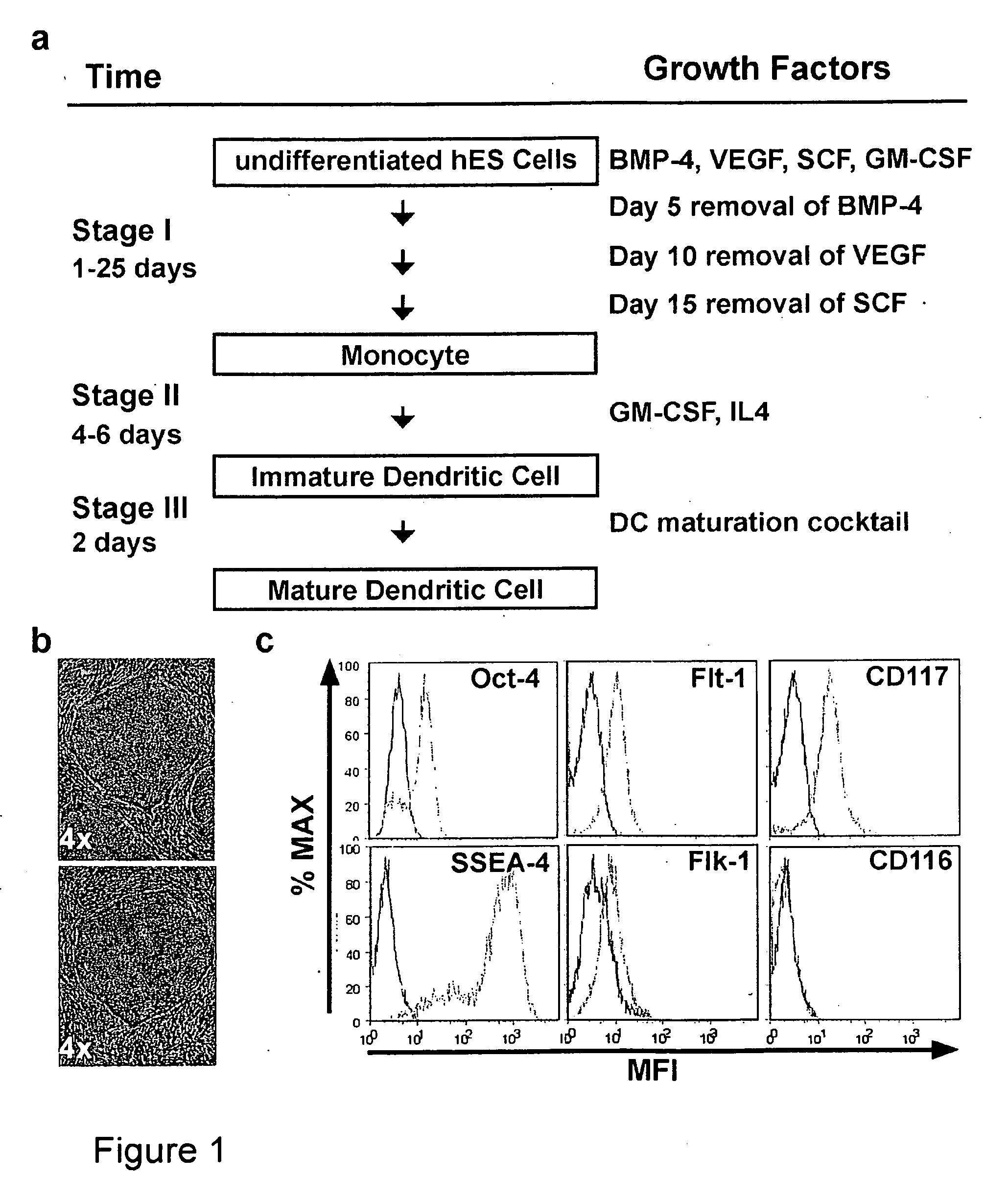
[0190]In this example pPS cells were differentiated into mDC by first culturing the pPS cells with a differentiation cocktail to obtain imDC and then further culturing the imDC with a maturation cocktail. The differentiation cocktail comprised exogenous cytokines which varied over the course of the experiment as the cells were differentiated to the imDC stage. (FIG. 1a). Human ES cell line H1 (Thomson et al., (1998) Science 282:1145) were cultured feeder free in defined serum-free media devoid of animal-derived products (Xu et al., (2001) Nat Biotechnol 19:971; Li et al., (2005) Biotechnol Bioeng 91:688) (FIG. 1b). The cells were also cultured stromal cell free throughout the differentiation and maturation protocol.
[0191]The hES cells were cultured under conditions permissive for forming embryoid bodies (EBs). Briefly, H1 cells were treated with collagenase D, (Invitrogen, Carlsbad, Calif.) rinsed once with 1...
example 2
Time Course Analysis of Differentiating Cell Cultures
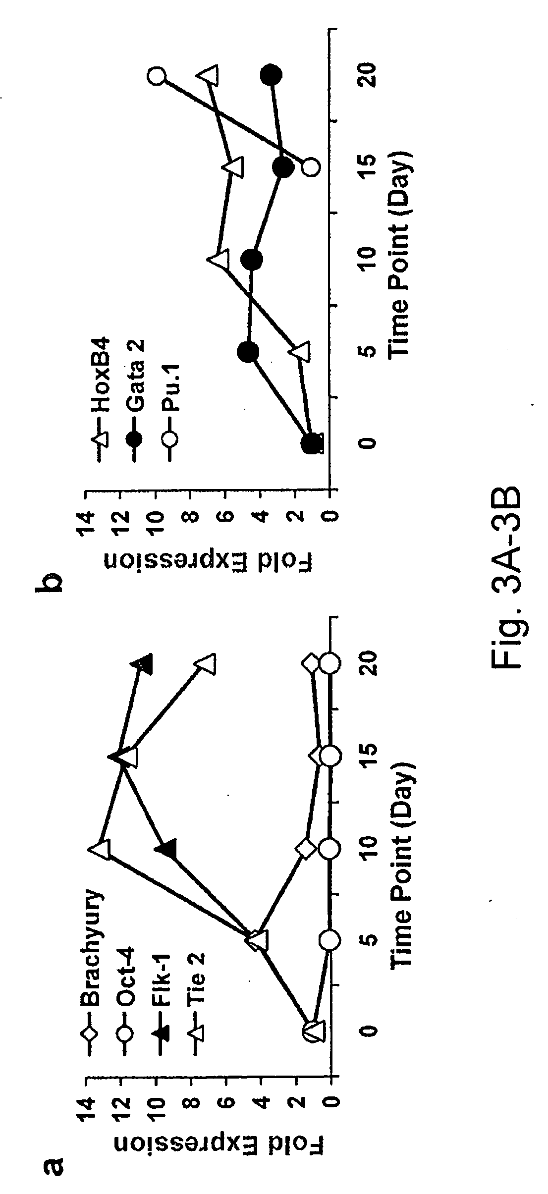
[0196]In order to characterize precursor cell populations arising during the process of differentiating pPS to imDC, the cell culture described in Example 1 was evaluated over time for transcription factor expression using RT PCR and real time quantitative PCR (as described below) and by flow cytometry (as described above) over time for expression of cell surface markers.
[0197]For real time quantitative PCR target cells were harvested and total RNA was isolated following the standard Qiagen RNeasy® Mini Prep protocol (Qiagen, Valencia, Calif.). Qiagen QiaShredder (Qiagen, Valencia, Calif.) was used to homogenize the lysate. Isolated RNAs were stored at −80° C. For cDNA synthesis, 1 μg RNA samples were treated with DNase (Ambion, Austin Tex.) to remove any genomic DNA impurities from the RNA prep. Reverse Transcriptase PCR(RT-PCR) was performed using Superscript II™ (Invitrogen, Carlsbad, Calif.) first strand synthesis system to ma...
example 3
Antigen Processing and Presentation
[0202]To test the ability of the hES derived imDC to process antigen, the fluorescent dye, DQ-OVA (Invitrogen, Carlsbad, Calif.), was dissolved at 1 mg / ml in PBS and added at 100 μg / ml to imDC derived from hES as described in Example 1. The protein was labeled with a pH insensitive BODIPY-F1 dye. The dye is self quenching when the protein is intact, but fluoresces bright green when the protein is denatured or undergoes proteolysis. The cells were incubated either at 37° C. or at 4° C. (as a control for background fluorescence) and washed 2× with flow buffer. Data was collected with FACSCalibur™ (Becton Dickinson, San Jose, Calif.) in FL1. The treated cells were found to fluoresce indicating that the protein had been proteolyzed by the imDC, while the control cells did not (FIG. 5a).
[0203]A functional assay was performed next to determine if DC made according to the method of Example 1 were able to stimulate antigen specific lymphocyte secretion of ...
PUM
| Property | Measurement | Unit |
|---|---|---|
| Fraction | aaaaa | aaaaa |
| Fraction | aaaaa | aaaaa |
| Fraction | aaaaa | aaaaa |
Abstract
Description
Claims
Application Information
 - R&D Engineer
- R&D Manager
- IP Professional
- Industry Leading Data Capabilities
- Powerful AI technology
- Patent DNA Extraction
Browse by: Latest US Patents, China's latest patents, Technical Efficacy Thesaurus, Application Domain, Technology Topic.
© 2024 PatSnap. All rights reserved.Legal|Privacy policy|Modern Slavery Act Transparency Statement|Sitemap