
Method and device for processing and presenting medical images

a technology for medical images and images, applied in image enhancement, image analysis, instruments, etc., can solve the problems that the magnification window does not necessarily move in a geometric consistent way or in accordance, and achieve the effect of facilitating the examination of such images, reducing the time to compare details, and more detailed looking at parts

Inactive Publication Date: 2009-10-15
IBBT +3
View PDF5 Cites 55 Cited by
  • Summary
  • Abstract
  • Description
  • Claims
  • Application Information

AI Technical Summary

Benefits of technology

[0035]By mapping regions of interest between the two images, it is possible to provide a medical expert with the interesting parts of the image upon request. In other words, a medical expert is able to assess all the important regions of an image without the need to look at the entire image. Of course, if the user or medical expert desires to do so, he is still able to inspect each part of the images to ensure that he did not miss anything.
[0056]Showing such information on top of the images, in the magnification windows or a separate window makes it easy for a medical expert to make a diagnosis or to determine how well treatment has an effect on a particular structure or disease. In addition, the medical expert no longer needs to perform measurements by hand, count the number of elements in a region etc. which reduces the time needed for the analysis
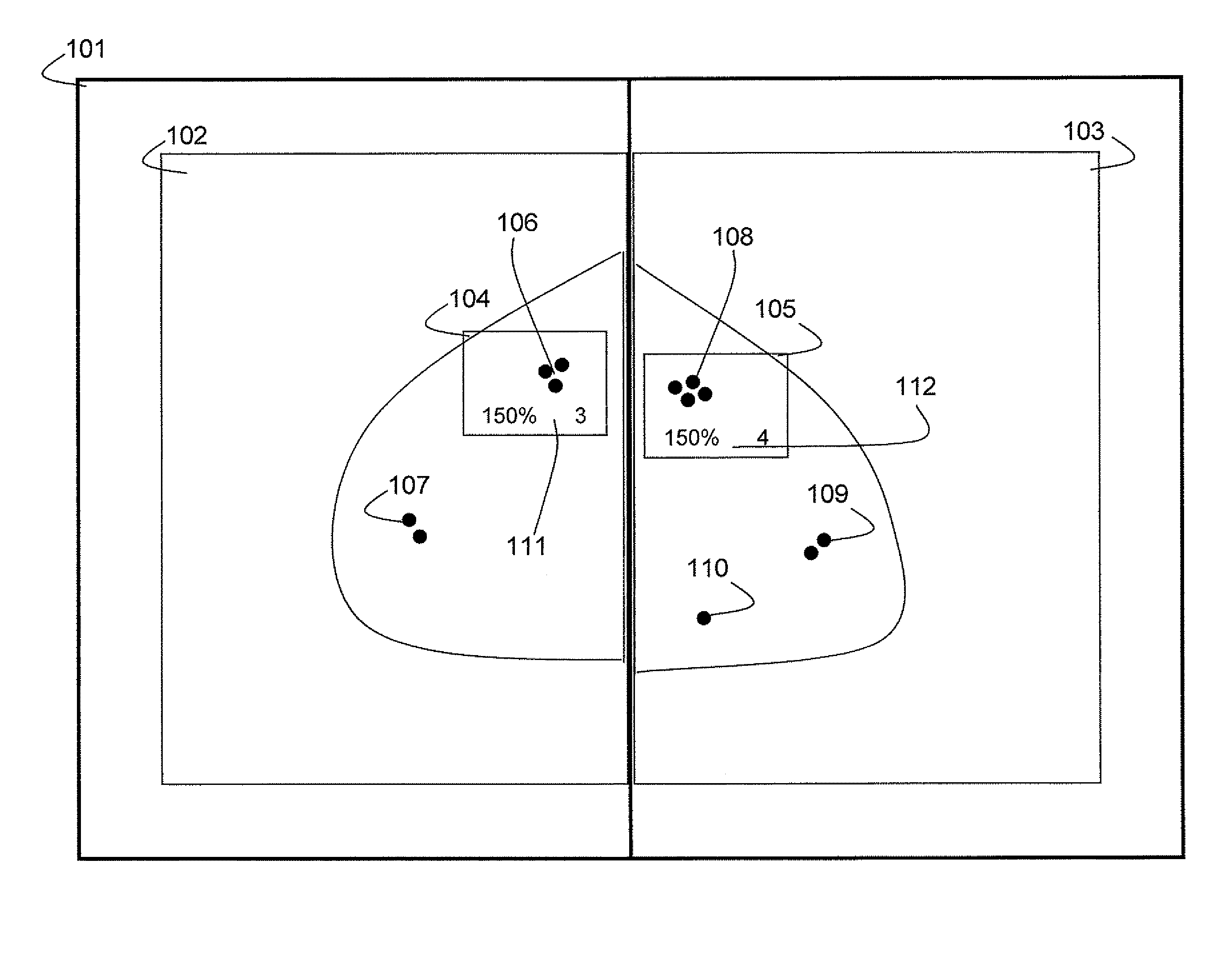
[0072]It is possible to present the registered images to a medical expert which provides the expert with a view on two images for comparison thereof. To facilitate the examination of such images, a magnification window is provided on both images. This way the expert is able to have a more detailed look at parts of an image without the need for additional tools such as a magnification glass. By presenting a magnification window on both images, the expert does not have to move the magnification window between the two images which reduces the time to compare details. However, images may be deformed during the registration phase which means that presenting registered images may result in incorrect or incomplete images being shown. A medical expert may therefore not be able to assess the contents of these images correctly which is unacceptable. It may therefore be advantageous to present the original images to the medical expert or a person looking at the images and present the magnification windows on these original images. Of course it may be possible for a user or a medical expert to select which images are displayed. It may even be possible to present an image in its original form and registered form if the user desires so.
[0081]The device presents corresponding regions to the user in a way similar as described with respect to the method above. Thus the device provides the same advantages of the pixel mapping or region-level correspondences as described above.

Problems solved by technology

However, compared to the prior art solutions, the magnification windows do not necessarily move in a geometric consistent way or in accordance with image data.

Method used


Image

  • Method and device for processing and presenting medical images
  • Method and device for processing and presenting medical images
  • Method and device for processing and presenting medical images

Examples


Embodiment Construction

)

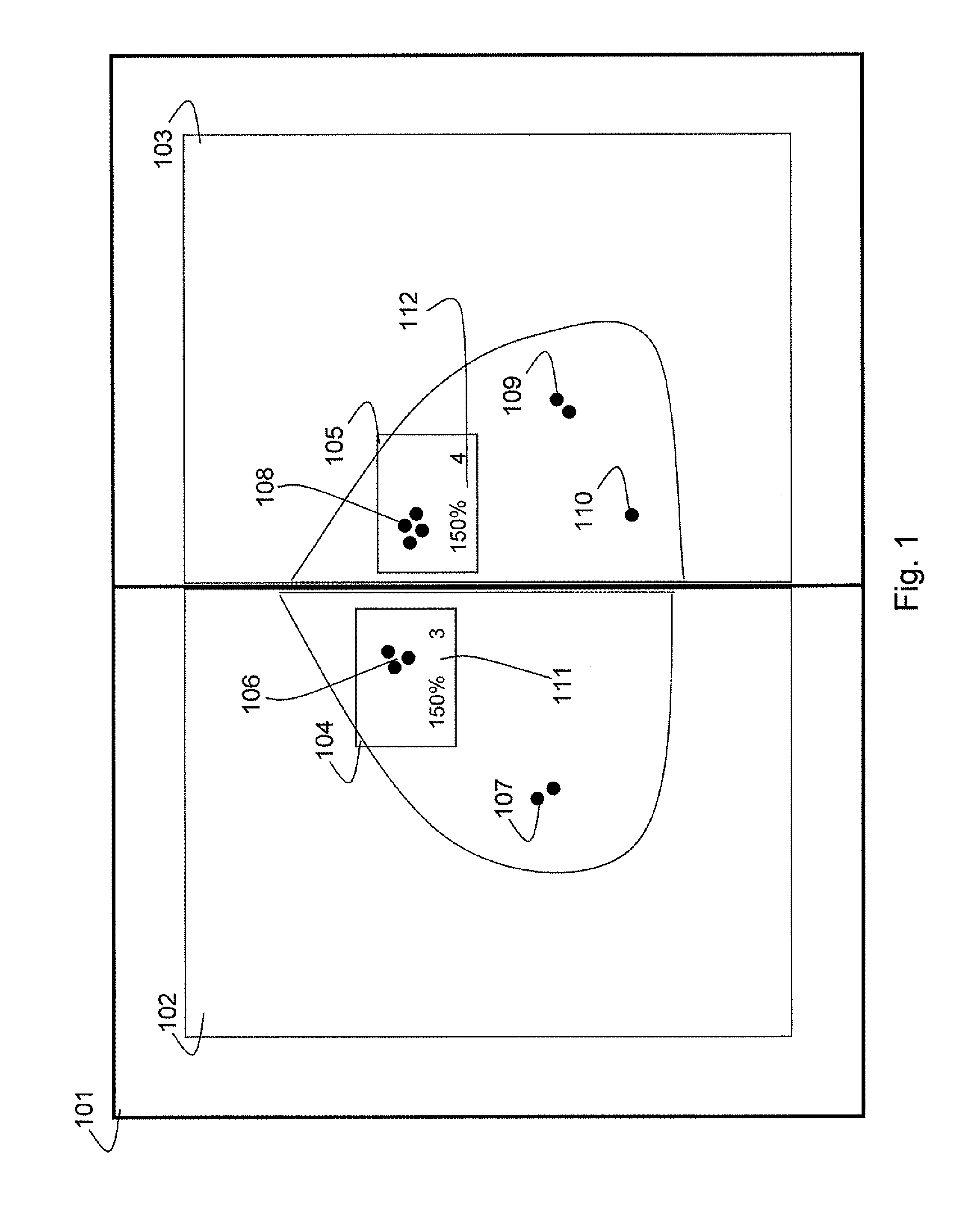
[0088]FIG. 1 illustrates a display 101 whereon two images 102 and 103 are presented. The image 102 and 103 are related to the right breast of a woman and show a medio-lateral oblique projection of the same breast. Image 102 is the current image which was taken recently whereas image 103 is the prior image which was taken at an earlier point in time, typically a year or two earlier. The display further presents a magnification window 104 on the current image 102 and a magnification window 105 on the prior image 103. Each of the images 102 and 103 in this particular example show a number of micro-calcifications 106-110. The current image 102 shows two locations with micro-calcifications 106 and 107. The prior image 103 shows three locations with micro-calcifications 108, 109 and 110.

[0089]In this example it is assumed that the current image 102 and the prior image 103 have been processed before being presented on the display 101. Thus the images 102 and 103 have gone through a regist...


PUM


Abstract

The present invention relates to a method for processing and presenting at least a first image (102) and a second image (103), these images being digital medical images. A first step in the method is performing image registration between the first image (102) and the second image (103) to generate a pixel-level mapping between both images. The registration takes into account a region-level correspondence between the first image (102) and the second image (103) A second step is presenting the first image (102) and the second image (103) simultaneously on a display (103). A third step is presenting a first magnification (104) of a region of interest (106) in the first image (102) and a second magnification (105) of a corresponding region (108) in the second image (103). The corresponding region (108) in the second image (103) being determined based on the pixel-level mapping.

Description

FIELD OF THE INVENTION[0001]The present invention generally relates to the presentation of medical images and more in particular to processing and presenting multiple images for diagnostics and computer assisted diagnostics (CAD).BACKGROUND OF THE INVENTION[0002]Medical or clinical images such as X-ray images, Computer Tomography (CT) scans, magnetic resonance imaging (MRI) etc. can be obtained by the use of specific devices and yield a specific type of images. The analysis of such images is generally carried out by a skilled person such as a radiologist or a doctor in a relevant field of medicine. The skilled person will typically look at the images and scan them for any irregularities which may provide a clue to the cause of pain or a disease. For instance looking at an X-ray image of an arm or a leg provides sufficient information to determine whether a bone is fractured. Similarly swellings in the tissue such as a tumour or other irregular structures in a particular part of the ...

Claims


Application Information

Patent Timeline
IPC IPC(8): G06K9/46
CPCG06T3/0075G06T7/0012G06T7/0034G06T2207/30068G06T2207/10072G06T2207/10116G06T7/401G06T7/35G06T7/41G06T3/147
Inventor TEMMERMANS, FREDERIKDEKLERCK, RUDISULIGA, MAREKBEHIELS, GERTDEWAELE, PIETBREUCQ, CATHERINEDE MEY, JOHAN
Owner IBBT
Learn More